Is Dopamine the Pleasure Chemical? Not Quite. 390 Fly Neurons, One Receptor, and What the Experiments Say
We counted the predicted dopamine neurons in a complete fruit-fly connectome, looked at dopamine bound to the human D1 receptor, and checked the 'pleasure chemical' label against the monkey and rat experiments behind it. Includes the four things we fixed before publishing.
This is the companion post to our Short Is dopamine the pleasure chemical? Not quite. The video is under a minute long. This post has the numbers, where each came from, and what we got wrong on the way.
한국어판: 도파민은 쾌락 물질일까? 꼭 그렇지는 않습니다
Educational content, not medical advice.
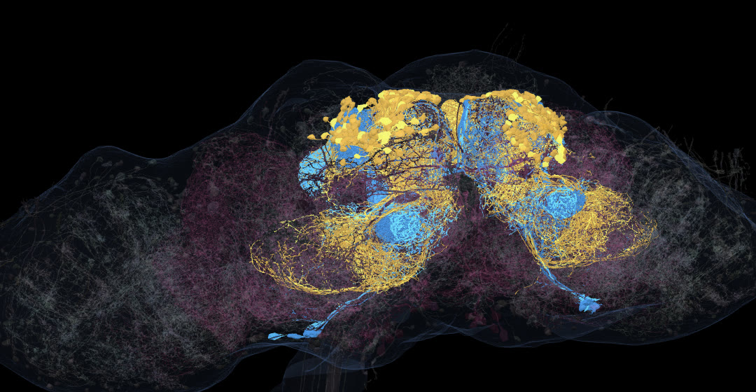 Gold: the 316 PAM neurons. Blue: the 12 PPL1 neurons of the mushroom-body types. Faint pink: the other 62 predicted dopamine neurons. Grey: 600 other neurons for scale. Real meshes from the Janelia male CNS connectome.
A familiar label
Dopamine is often called the pleasure chemical. The label is short and easy to remember. According to the experiments that made dopamine famous, it is also not quite right.
We checked it three ways: counting dopamine neurons in a complete fruit-fly wiring map, looking at dopamine itself in a human receptor, and going back to the monkey and rat studies behind the "not pleasure" argument. One thing up front: the wiring map cannot tell you what dopamine feels like. It shows how few dopamine neurons there are and where their output goes.
Finding dopamine in a whole nervous system
The Janelia male CNS connectome maps the entire central nervous system of one male fruit fly: 166,700 neurons across the brain and nerve cord (Berg et al., Cell 2026). Each neuron carries a predicted neurotransmitter: not a chemical measurement, but a machine-learning prediction made from electron-microscope images of its synapses.
How good is it for dopamine? The male CNS paper shows its accuracy only in a figure. For the published method used on the FlyWire and hemibrain connectomes, Eckstein et al. (Cell 2024) found dopamine predicted correctly for about 90% of known dopaminergic cell types in FlyWire and 85% in the hemibrain. It also has a known failure: it wrongly labels Kenyon cells, the main neurons of the fly's learning centre, as dopaminergic. That does not affect our count, because no Kenyon cells are in our set.
392 neurons in the male CNS have the consensus label "dopamine". We removed two of them, the DPM neurons, because earlier experiments tie them to other transmitters: serotonin (Lee et al., PNAS 2011) and GABA (Haynes et al., eLife 2015). That leaves 390.
| Group | Neurons (both sides) | What we know about them |
|---|---|---|
| PAM (15 types, PAM01–PAM15) | 316 | Generally signal reward |
| PPL1, mushroom-body types (PPL101–PPL106) | 12 | Generally signal punishment |
| PPL1, other types (PPL107, PPL108) | 4 | Not mushroom-body neurons |
| PPM1 | 12 | |
| PPL2 | 6 | |
| Other named types (fan-shaped body, visual centrifugal LoVC and others) | 40 | |
| Total | 390 |
All 390 are named neurons with a cell type, not unassigned fragments. That is about 1 in 420 of the 164,620 neurons with a transmitter prediction.
How does that compare with older counts?
Before connectomes, fly dopamine neurons were counted under the light microscope with antibodies against tyrosine hydroxylase (TH), an enzyme needed to make dopamine. Mao and Davis (2009) counted about 282 TH-positive neurons per brain in the protocerebrum (the central brain, not the optic lobes), including about 100 PAM neurons per hemisphere. Other methods give 98 per hemisphere (split-GAL4) or 115–135 (dopamine immunostaining) (Aso et al. 2014, citing Liu et al. 2012).
Our 316 PAM neurons across both sides is about 158 per side, higher than any of the light-microscopy counts. Two things could contribute: our labels are predictions and can include neurons that are not truly dopaminergic, and an electron-microscope reconstruction can catch faint cells an antibody stain misses. Our data cannot tell which matters more. The earlier hemibrain connectome also found more than light microscopy did: Li et al. (2020) report 158 dopamine neurons innervating the mushroom body, about 150 of them PAM.
The PPL1 number lines up better. Li et al. define six PPL1 types that go to the mushroom body (PPL101–PPL106), with "only a single cell per PPL1-DAN cell type in a hemisphere". Six types, one cell each, on two sides: 12. That is exactly what we found.
For people, the comparison is rough. Pakkenberg et al. (1991) counted an average of 550,000 pigmented neurons in the substantia nigra of seven elderly control brains. Against the 86.1 billion neurons of an adult human brain (Azevedo et al. 2009), that is about 1 in 157,000. The methods differ, the human number covers one dopamine region only, and the abstract does not say whether it counts one side or both. Read "1 in 420 versus about 1 in 157,000" as a rough illustration that dopamine neurons are rare in both, not as a precise ratio.
Where the output goes
Together the 390 predicted dopamine neurons make 803,309 output synapses, each one a pre-to-post connection counted on the receiving side. The PAM neurons make 271,752 of them, and 173,789 of those land on Kenyon cells: 64%, nearly two-thirds. That is a lower bound for the learning centre, because we counted only Kenyon-cell targets, not any other neurons there.
The four PPL107 and PPL108 neurons are a contrast: 36,970 output synapses between them, only 14 to Kenyon cells.
One more number needs care. All 392 dopamine-labelled bodies together connect to 295,018 distinct partner bodies, but only 39,301 of those (about 13%) are named neurons. The rest are fragments: real tissue the reconstruction never joined to a whole cell. In an earlier post, counting fragments as neurons nearly quadrupled a number, so we now report reach in named neurons only.
Reward and punishment, generally
What do PAM and PPL1 do? Here the evidence is experimental, not anatomical. Liu et al. (Nature 2012) found that "a group of dopamine neurons in the protocerebral anterior medial (PAM) cluster signals sugar reward". Burke et al. (Nature 2012) showed that activating PAM subsets directly "can substitute for sugar to form appetitive memory". On the other side, Claridge-Chang et al. (Cell 2009) "mapped the origin of aversive reinforcement signals to the PPL1 cluster of 12 dopaminergic cells".
Their 12 cells are one PPL1 cluster on one side, not all of which reach the mushroom body. Our 12 are the mushroom-body types on both sides. The numbers match by coincidence.
The connectome paper words it carefully: PPL1 neurons "generally convey punishment, whereas PAM DANs … generally convey reward" (Li et al. 2020). The word "generally" matters. Some PAM types can produce aversive memories (Aso et al. 2012), and a single dopamine cell type can write an appetitive or an aversive memory depending on when it fires relative to an odour (Aso & Rubin 2016). That is why the video says "mainly reward" and "mostly punishment".
Even in a fly, then, dopamine is not simply "the good feeling". The same chemical teaches the learning centre about both reward and punishment. (For an older, broader overview of how addictive drugs act on the human reward system, see our Neuroscience of Addiction Explained.)
Dopamine itself, in its receptor
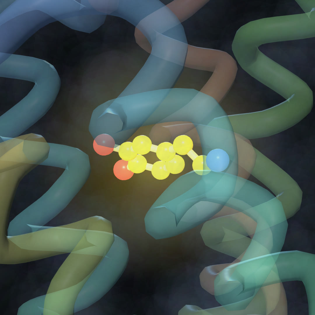 Dopamine bound in the human D1 receptor. Real cryo-EM structure, PDB 7F0T.
Dopamine acts by binding receptors on the receiving cell. PDB entry 7F0T shows the human D1 dopamine receptor in complex with a mini-Gs protein and dopamine as the only bound ligand, solved by cryo-electron microscopy at 3.1 Å (Teng et al., Sci Adv 2022). Several other dopamine-bound D1 structures also contain a modulating drug, which is why we chose this one. It shows where dopamine sits; like the connectome, it says nothing about pleasure. For that we need behaviour.
Not pleasure: what monkeys and rats showed
Surprise, not reward itself. Drawing on recordings in monkeys, Schultz, Dayan and Montague described dopamine neurons "whose fluctuating output apparently signals changes or errors in the predictions of future salient and rewarding events" (Schultz, Dayan & Montague, Science 1997). A later review summarises the pattern in three parts: most midbrain dopamine neurons "are activated by more reward than predicted …, remain at baseline activity for fully predicted rewards, and show depressed activity with less reward than predicted" (Schultz 2016). A fully expected reward does not make them fire more, which is hard to square with a simple pleasure signal.
Caveats: these neurons also respond to cues that predict reward, and not all of them behave alike. In monkeys, Matsumoto and Hikosaka (2009) found that "a greater number of dopamine neurons were excited by both" reward-predicting and airpuff-predicting stimuli.
Wanting, not liking. Berridge and Robinson (1998) depleted dopamine in the nucleus accumbens and neostriatum of rats "by up to 99%". The rats still showed "normal hedonic reaction patterns to sucrose vs. quinine": the reactions that signal liking a sweet taste and disliking a bitter one. Their conclusion was that "dopamine systems are necessary for 'wanting' incentives, but not for 'liking' them".
This is a hypothesis, and it is still debated. Others describe dopamine as "a critical modulator of both learning and motivation" (Berke 2018). Either way, dopamine looks more like a signal for learning and for going after things than the feeling of enjoyment itself, which is why the video says it "seems to" drive wanting.
For how drugs act on this reward system, see our earlier explainer, Neuroscience of Addiction Explained.
What we got wrong (and fixed before publishing)
An adversarial check before rendering, a second pass that tries to break each number and claim, caught four problems.
- Sixteen punishment neurons became twelve. The first draft counted every PPL1-named neuron: 16. But PPL107 and PPL108 (4 cells) are not mushroom-body types (Scheffer et al. 2020; Li et al. 2020), and in our data they send almost nothing to Kenyon cells (14 of 36,970). The punishment role is a mushroom-body role, so the number is 12. In the figure they are pink, with the "other" group.
- The title said too much. The first title was "Real brain-map data says no". But the "not pleasure" evidence comes from rats and monkeys; the connectome shows only counts and wiring. The title became "Not quite".
- Claims without species or hedging. The first draft stated prediction error and wanting-versus-liking as plain facts. The recordings are from monkeys, the depletion study is in rats, and wanting-versus-liking is a debated hypothesis. The captions now say "In monkeys" and "seems to".
- Two neurons removed on purpose. The prediction calls the two DPM neurons dopaminergic, but experiments point to serotonin and GABA. Where a prediction conflicts with experiments, we follow the experiments: 390, not 392. (The partner count above still covers all 392.)
What this does not show
- The transmitter labels in the fly connectome are predictions, not measurements of dopamine in each cell.
- The connectome shows connections, not function. The reward and punishment roles come from behavioural experiments.
- The fly and human comparison mixes different counting methods, regions and denominators.
- The prediction-error and wanting-versus-liking evidence is from monkeys and rats. How far it carries over to human experience is a separate question.
Code and data
All files are in the shoo99/science-shorts repository.
- Fly: Janelia FlyEM male CNS v1.0 (CC BY). Predicted transmitter (
consensusNt:dopamine) and cell types from the public segment properties; output synapses per neuron from the public synapse table, counted as pre-to-post connections. Kenyon-cell targets are partners whose type starts with KC; named partners are partners with a cell type. Counts:verify/fly-dopamine.js→verify/out/fly-dopamine.json. Figure scene:data/fetch-fly-da.js(DPM excluded). - Structure: PDB 7F0T (human D1 receptor, mini-Gs, dopamine).
- Source wording for every claim above:
verify/out/dopamine-factbase.md. - The figure renders real meshes; the light pulses in the video are illustrative.
References
- Berg S., Beckett I.R. et al. (2026). Sexual dimorphism in the complete Drosophila male central nervous system connectome. Cell 189:5504–5526. doi:10.1016/j.cell.2026.08.015
- Eckstein N., Bates A.S. et al. (2024). Neurotransmitter classification from electron microscopy images at synaptic sites in Drosophila melanogaster. Cell 187:2574–2594. doi:10.1016/j.cell.2024.03.016
- Lee P.T. et al. (2011). Serotonin–mushroom body circuit modulating the formation of anesthesia-resistant memory in Drosophila. PNAS 108:13794–13799. doi:10.1073/pnas.1019483108
- Haynes P.R., Christmann B.L., Griffith L.C. (2015). A single pair of neurons links sleep to memory consolidation in Drosophila melanogaster. eLife 4:e03868. doi:10.7554/eLife.03868
- Mao Z., Davis R.L. (2009). Eight different types of dopaminergic neurons innervate the Drosophila mushroom body neuropil. Front Neural Circuits 3:5. doi:10.3389/neuro.04.005.2009
- Aso Y. et al. (2014). The neuronal architecture of the mushroom body provides a logic for associative learning. eLife 3:e04577. doi:10.7554/eLife.04577
- Liu C. et al. (2012). A subset of dopamine neurons signals reward for odour memory in Drosophila. Nature 488:512–516. doi:10.1038/nature11304
- Burke C.J. et al. (2012). Layered reward signalling through octopamine and dopamine in Drosophila. Nature 492:433–437. doi:10.1038/nature11614
- Claridge-Chang A. et al. (2009). Writing memories with light-addressable reinforcement circuitry. Cell 139:405–415. doi:10.1016/j.cell.2009.08.034
- Li F. et al. (2020). The connectome of the adult Drosophila mushroom body provides insights into function. eLife 9:e62576. doi:10.7554/eLife.62576
- Scheffer L.K. et al. (2020). A connectome and analysis of the adult Drosophila central brain. eLife 9:e57443. doi:10.7554/eLife.57443
- Aso Y. et al. (2012). Three dopamine pathways induce aversive odor memories with different stability. PLoS Genet 8:e1002768. doi:10.1371/journal.pgen.1002768
- Aso Y., Rubin G.M. (2016). Dopaminergic neurons write and update memories with cell-type-specific rules. eLife 5:e16135. doi:10.7554/eLife.16135
- Pakkenberg B. et al. (1991). The absolute number of nerve cells in substantia nigra in normal subjects and in patients with Parkinson's disease estimated with an unbiased stereological method. J Neurol Neurosurg Psychiatry 54:30–33. doi:10.1136/jnnp.54.1.30
- Azevedo F.A. et al. (2009). Equal numbers of neuronal and nonneuronal cells make the human brain an isometrically scaled-up primate brain. J Comp Neurol 513:532–541. doi:10.1002/cne.21974
- Teng X. et al. (2022). Structural insights into G protein activation by D1 dopamine receptor. Sci Adv 8:eabo4158. doi:10.1126/sciadv.abo4158
- Schultz W., Dayan P., Montague P.R. (1997). A neural substrate of prediction and reward. Science 275:1593–1599. doi:10.1126/science.275.5306.1593
- Schultz W. (2016). Dopamine reward prediction error coding. Dialogues Clin Neurosci 18:23–32. doi:10.31887/DCNS.2016.18.1/wschultz
- Matsumoto M., Hikosaka O. (2009). Two types of dopamine neuron distinctly convey positive and negative motivational signals. Nature 459:837–841. doi:10.1038/nature08028
- Berridge K.C., Robinson T.E. (1998). What is the role of dopamine in reward: hedonic impact, reward learning, or incentive salience? Brain Res Rev 28:309–369. doi:10.1016/S0165-0173(98)00019-8
- Berke J.D. (2018). What does dopamine mean? Nat Neurosci 21:787–793. doi:10.1038/s41593-018-0152-y
More from Inside the Connectome: Why do some allergy pills make you sleepy? · Human vs mouse vs fly: how we measured five rounds · Same lock, different effects: LSD and lisuride
Author — the operator of kbrain-map.org, a researcher who analyses multi-omics (proteomics) data. The measurements behind the Inside the Connectome videos and this post were made directly on the public connectome and protein-structure data.
A note from the operator — The video is under a minute long, so it could not show everything behind the numbers. If you find a mistake, please tell us: we will fix the post and pin a correction under the video.
Related
Why Do Some Allergy Pills Make You Sleepy? Eight Histamine Neurons, One Receptor Pocket, and a Door
Oct 20 · 11 min read
NeuroscienceHuman vs Mouse vs Fly: How We Measured Five Rounds of Brain-Cell Comparisons (and What We Got Wrong First)
Oct 16 · 15 min read
NeuroscienceHow Close Are Neurons to Blood Vessels? We Measured 10,441 of Them in a Real Human Brain Sample
Oct 30 · 6 min read
NeuroscienceWhich H01 Is It? Two Public Synapse Releases, Only One Matches the Paper — and How We Found Out the Hard Way
Oct 19 · 8 min read