How Close Are Neurons to Blood Vessels? We Measured 10,441 of Them in a Real Human Brain Sample
In about one cubic millimetre of human cortex mapped with an electron microscope (H01), 98% of neurons sit within 50 µm of a blood vessel (median 23 µm). How we measured it, why neurons stay so close, and what the number does not tell you.
This is the companion post to our Short How close is every neuron to a blood vessel?. The number is also on our Numbers page, with the method and the caveats in one card.
The short answer
In a piece of real human cortex, about one cubic millimetre, 98% of neurons have a blood vessel within 50 micrometres of the centre of their cell body. The median distance is 23 µm. For scale, a human hair is roughly 50–100 µm wide, and a neuron's cell body is about 10–20 µm across. So most neurons sit about one cell-width away from a vessel.
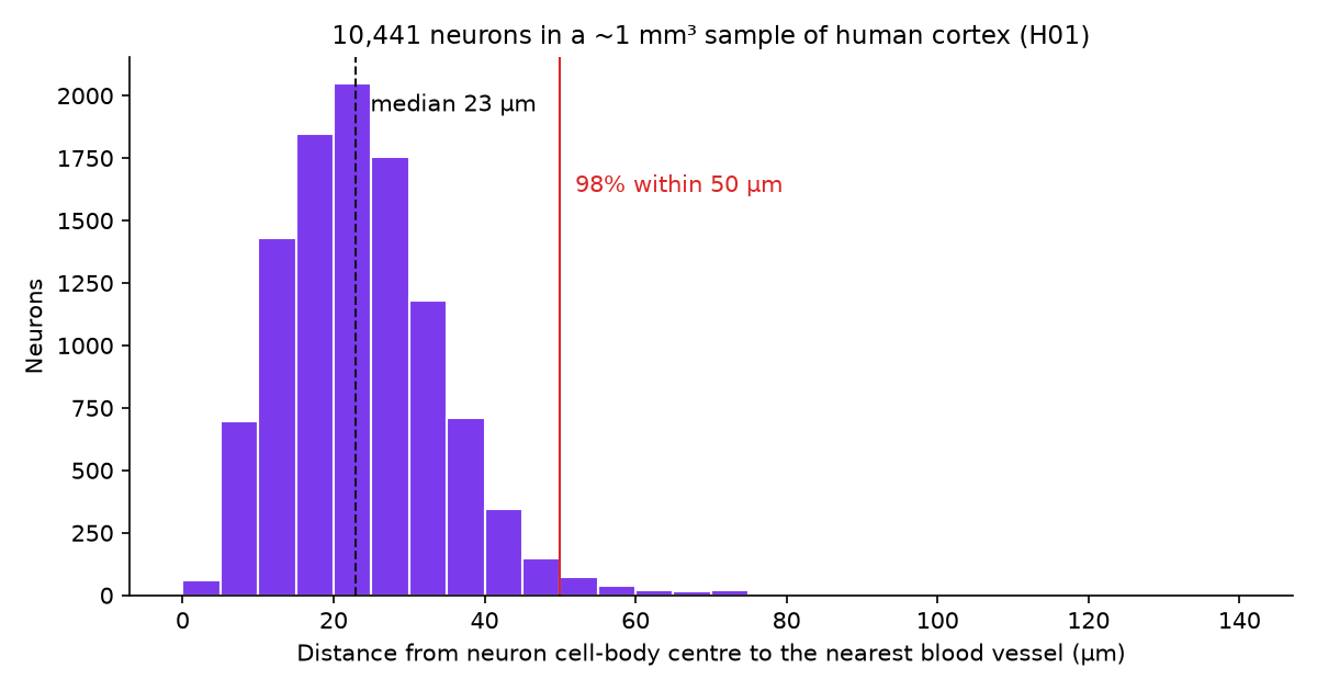
Distance from each neuron's cell-body centre to the nearest point on a blood-vessel surface. 10,441 neurons, H01. Our measurement.
The map we used
H01 is a fragment of human temporal cortex, removed during epilepsy surgery, imaged with an electron microscope at nanometre resolution and reconstructed in 3D (Shapson-Coe et al. 2024, Science). It is about 3 × 2 mm wide and 0.17 mm thick, and it is public under CC BY 4.0.
Two parts of the release did all the work:
- Blood vessels. The H01 team's gallery view "blood_vessels" ("Vasculature within the tissue rendered in 3D") lists 1,479 segments of the
blood_vessels_segmentedlayer. 1,477 of them have a 3D surface mesh; two have none. We downloaded every mesh at the finest level of detail: 1.34 million triangles. - Cell bodies. The release's
somas.csvtable gives the position and type of every cell body. 16,087 are labelled as neurons (pyramidal, interneuron, spiny atypical, spiny stellate, unclassified neuron), exactly the number in the paper's Table S5.
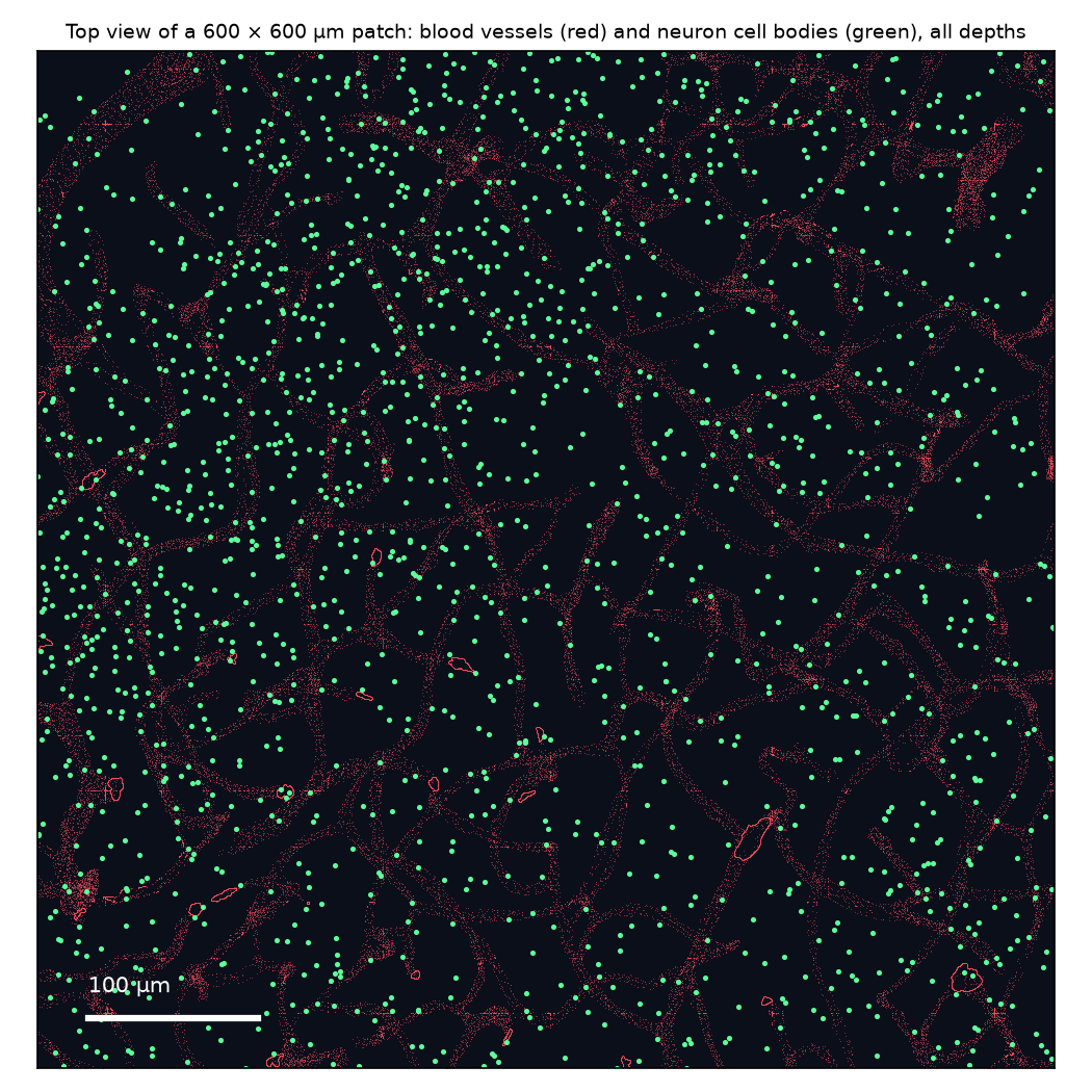
A 600 × 600 µm patch seen from above, all 170 µm of depth flattened. Red: vessel surfaces. Green: neuron cell bodies. Because depth is flattened, a neuron that looks far from a vessel here may have one just above or below it.
How we measured it
- Leave out the edges. A neuron near the edge of the sample may have its nearest vessel just outside the tissue we have. We kept only neurons at least 50 µm inside the sample in the two wide directions and 30 µm inside in the thin direction: 10,441 of the 16,087.
- Nearest vessel. For each of these neurons we took the distance from its cell-body centre to the nearest vertex of any vessel mesh. The mesh vertices are about 1.6 µm apart, so this slightly over-estimates the true distance to the vessel wall.
- Summarise. Median 22.9 µm. 39% are within 20 µm, 75% within 30 µm, 98% within 50 µm. The farthest is 141 µm.
We measured the same thing for glia (astrocytes, oligodendrocytes, microglia and their precursors): 92% sit within 50 µm of a vessel, a little less than neurons.
Why neurons stay so close
The brain is expensive. It is about 2% of body weight but uses about 20% of the body's oxygen and calories (Raichle & Gusnard 2002, PNAS). It also keeps little fuel in reserve, so it depends on a steady supply from the blood. Oxygen and glucose leave the smallest vessels, the capillaries, and have to diffuse through tissue to reach cells; diffusion is fast over a few tens of micrometres and slow beyond that. A dense web of capillaries, with every neuron close to one, is the brain's answer.
How this compares with other measurements
The best-known number comes from mouse cortex. Tsai et al. (2009, J Neurosci) mapped every nucleus and vessel in thick slabs of mouse neocortex and found that "the distance from the neuronal nuclei to the nearest microvessel is 14.6 ± 1.0 μm", about 15 µm.
Our median of 23 µm is larger, but the two numbers are not measured the same way, so we do not read much into the gap:
- Tsai and colleagues measured to the centre line of the vessel; we measured to the vessel surface, which should make our numbers smaller, not larger.
- Their vessels were filled with a fluorescent gel pumped through the circulation; ours come from an automated segmentation of electron-microscope images, which may miss some capillaries. Missing capillaries would make our distances too large.
- It is a different species, a different brain area, and tissue from epilepsy surgery.
So treat 23 µm as an upper bound for this sample, and "within about two cell-widths" as the safe summary.
What this does not show
- Which vessels. We did not separate capillaries from larger arterioles and venules, so this is "any vessel", not "any capillary".
- Blood flow. A vessel nearby is not the same as a vessel that is carrying blood to that neuron at a given moment.
- One sample. One person, one area of cortex, removed during epilepsy surgery. Whether it is typical of healthy cortex is an open question that the H01 authors raise themselves.
- Cell-body centres, not whole cells. Neurons are mostly dendrites and axons, which spread hundreds of micrometres; the cell body is the only point we measured from.
Code and data
- Vessel meshes: H01
blood_vessels_segmented, finest level of detail, fetched by segment ID from the gallery view "blood_vessels". - Cell bodies: H01
somas.csv(positions and cell types). - Distances: nearest-neighbour search (SciPy
cKDTree) from each soma centre to all vessel-mesh vertices, after the edge filter above. The two figures are generated by the same script.
References
- Shapson-Coe A, Januszewski M, Berger DR, et al. A petavoxel fragment of human cerebral cortex reconstructed at nanoscale resolution. Science. 2024;384:eadk4858. doi:10.1126/science.adk4858
- Raichle ME, Gusnard DA. Appraising the brain's energy budget. PNAS. 2002;99:10237–10239. doi:10.1073/pnas.172399499
- Tsai PS, Kaufhold JP, Blinder P, et al. Correlations of neuronal and microvascular densities in murine cortex revealed by direct counting and colocalization of nuclei and vessels. J Neurosci. 2009;29:14553–14570. doi:10.1523/JNEUROSCI.3287-09.2009
Related
Which H01 Is It? Two Public Synapse Releases, Only One Matches the Paper — and How We Found Out the Hard Way
Oct 19 · 8 min read
NeuroscienceWe Followed One Neuron Through a Map of the Human Brain — Methods, Numbers, and What We Got Wrong
Oct 1 · 9 min read
NeuroscienceHuman vs Mouse vs Fly: How We Measured Five Rounds of Brain-Cell Comparisons (and What We Got Wrong First)
Oct 16 · 15 min read
NeuroscienceIs Dopamine the Pleasure Chemical? Not Quite. 390 Fly Neurons, One Receptor, and What the Experiments Say
Oct 27 · 13 min read