Why Do Some Allergy Pills Make You Sleepy? Eight Histamine Neurons, One Receptor Pocket, and a Door
We counted the histamine neurons in a complete fruit-fly connectome, overlaid two real antihistamine structures on the human H1 receptor, and followed the evidence for why only some allergy pills make you drowsy. Includes the two counting mistakes we caught before publishing.
This is the companion post to our Short Why do some allergy pills make you sleepy?. The video has 46 seconds. This post has the numbers, where each came from, and what we got wrong on the way.
한국어판: 비염약은 왜 어떤 건 졸리고 어떤 건 안 졸릴까?
Educational content, not medical advice. Questions about your own medicines belong with a doctor or pharmacist.
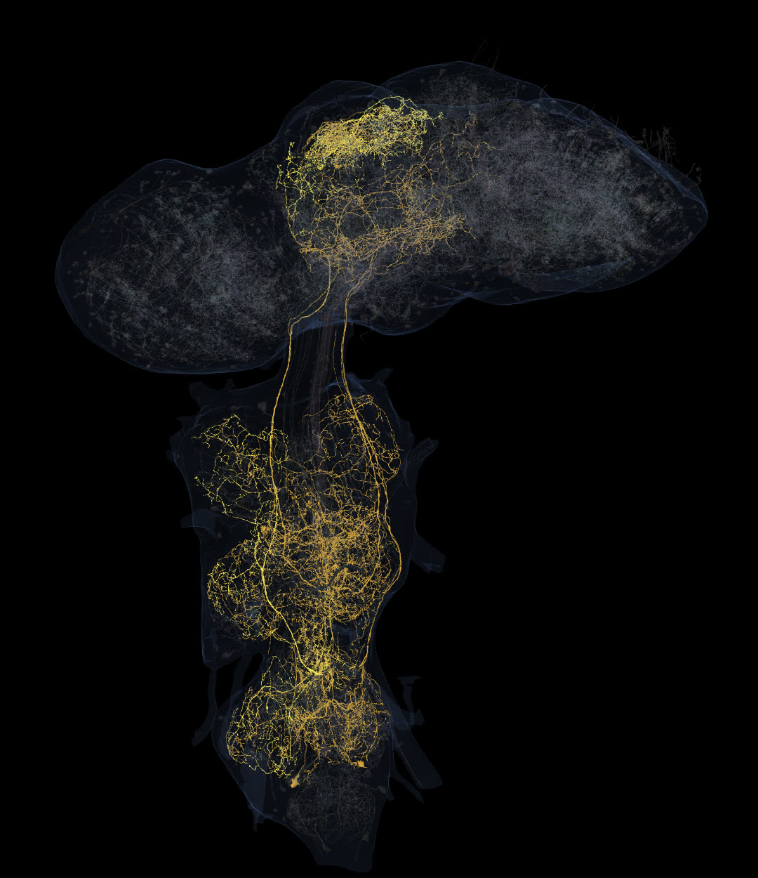 Gold: the eight named neurons outside the visual system that the fly connectome predicts use histamine. Grey: 600 other neurons for scale. Real meshes from the Janelia male CNS connectome.
A familiar puzzle
Take an old-fashioned allergy pill and you may want a nap. Take a newer one for the same sneezing and, for most people, nothing much happens to your alertness. Both are called antihistamines. Both are designed to block the same receptor. So why does only one of them reach your sense of being awake?
The usual answer is two sentences long: histamine keeps you awake, and the older drugs get into the brain. We wanted to see what that looks like in real data, and to check each half of the sentence against a primary source. We used three kinds of evidence: a complete wiring map of a fruit fly's nervous system, published structures of the human histamine receptor with the drugs bound, and the papers behind each claim.
Looking for histamine in a whole nervous system
The Janelia male CNS connectome maps the entire central nervous system of one male fruit fly: 166,700 neurons spanning the brain and nerve cord, all proofread (Berg et al., Cell 2026). Each neuron carries a predicted neurotransmitter, a consensus of machine-learning predictions made from the electron-microscope images. We read those labels directly from the public release.
5,910 neurons are predicted to use histamine. That sounded like a lot until we sorted them by where they sit. 4,114 are photoreceptors and other eye cells, which is expected: fly photoreceptors signal with histamine. Another 1,782 are neurons inside the optic lobes, the fly's visual processing centres, almost all of one type called T1. Outside the visual system, very few remain.
| Group | Neurons predicted to use histamine |
|---|---|
| Photoreceptors (eye, including the eyelet) | 4,114 |
| Optic-lobe neurons (1,777 of them T1) | 1,782 |
| Named neurons outside the visual system | 8 |
| Untyped partial bodies | 6 |
A caveat on these counts (added before publishing). They are machine predictions, and for histamine outside the eye they miss a lot. Antibody staining finds about 20 histamine neurons in the fly brain besides the photoreceptors ("approximately 12 neurons in each brain hemisphere", Pollack & Hofbauer, Cell Tissue Res 1991; 18 protocerebral plus 2 maxillary–labial neurons in later counts by Nässel), and about 18 more in the nerve cord (Nässel, Pirvola & Panula, J Comp Neurol 1990). Of our eight, only two (the GNG043 pair) sit in the brain proper. Histamine-using mechanosensory neurons are also known (Buchner et al. 1993), and none is predicted as histamine in the release. So read "eight" as "eight that the prediction flags", not as the fly's full histamine system.
The eight named neurons come in four pairs, one on each side: two in the gnathal ganglia at the base of the brain (type GNG043), four ascending neurons that run from the nerve cord up into the brain (AN27X008 and AN27X004), and two neurons inside the nerve cord (IN27X004). Between them they make 49,473 output synapses. Here an output synapse means a receiving contact: in flies a single release site (a T-bar) is usually contacted by several receiving neurites, so neuron 12230's 7,252 output synapses come from 1,928 release sites.
| Neuron (body ID) | Type | Output synapses | Partner bodies |
|---|---|---|---|
| 12230 | GNG043 | 7,252 | 4,555 |
| 12808 | GNG043 | 7,197 | 4,535 |
| 21783 | AN27X008 | 8,505 | 5,211 |
| 556586 | AN27X008 | 8,311 | 5,219 |
| 331721 | AN27X004 | 5,445 | 4,018 |
| 526576 | AN27X004 | 5,044 | 3,717 |
| 800415 | IN27X004 | 3,899 | 2,610 |
| 803748 | IN27X004 | 3,820 | 2,524 |
Counted once each, the partners of these eight include 7,976 annotated neurons. That is the number we used in the video: eight cells, reaching about eight thousand.
Two numbers we had to take back
Our first count was wrong twice, and both mistakes made the story sound better than it is.
First, we said fourteen neurons, not eight. The extra six were histamine-tagged bodies with no type, no name and no superclass. Their meshes have 717 to 2,642 vertices, against 26,000 to 49,000 for the named neurons, and they make 142 to 665 outputs each, against 3,800 to 8,500. They look like pieces of neurons that were never joined to a whole cell, so we stopped calling them neurons and removed them from the figure.
Second, we said these cells reach "over 30,000 neurons". That was the number of distinct partner bodies, 30,621 for all fourteen. But only 8,003 of those bodies are annotated neurons. The other 22,618 are unidentified fragments: real pieces of tissue the reconstruction never assigned to a cell. Counting them as neurons inflated the reach almost fourfold. Both corrections came from an adversarial check we run on every script before rendering, where a second pass tries to break each number.
What histamine does in a fly
The connectome tells us who is wired to whom. It does not tell us what histamine does. For that we went to experiments. In Oh et al. (2013, PLoS One), flies carrying weakened versions of the enzyme that makes histamine (histidine decarboxylase) slept longer than normal flies. Histamine, in other words, is a wake-promoting signal in flies too.
There is an important limit here. Those mutant flies have less histamine everywhere, including in their eyes, and the paper traces the wake effect through a histamine receptor on clock neurons. Nothing we measured shows that the eight gold neurons are the ones keeping a fly awake. The fly part of the story shows that the same chemical is used, and how few neurons outside the visual system use it, not which circuit does the waking.
The receptors are also different. A fly's histamine receptors (HisCl1 and Ort) are histamine-gated chloride channels. Ours are a different family of proteins. So the fly does not explain the drug. It explains the signal.
Histamine in your brain
In mammals, histamine-making neurons sit in a single small cluster, the tuberomamillary nucleus of the posterior hypothalamus, and send their axons all over the central nervous system. They are active only during waking and help maintain wakefulness and attention (Haas, Sergeeva and Selbach, Physiol Rev 2008). There is a pattern worth noticing: in both animals, a small number of histamine neurons reaches a large part of the nervous system.
One of the receptors that picks up this signal in the brain is the histamine H1 receptor. It is also the receptor that allergy pills are designed to block in the nose, eyes and skin.
Same lock
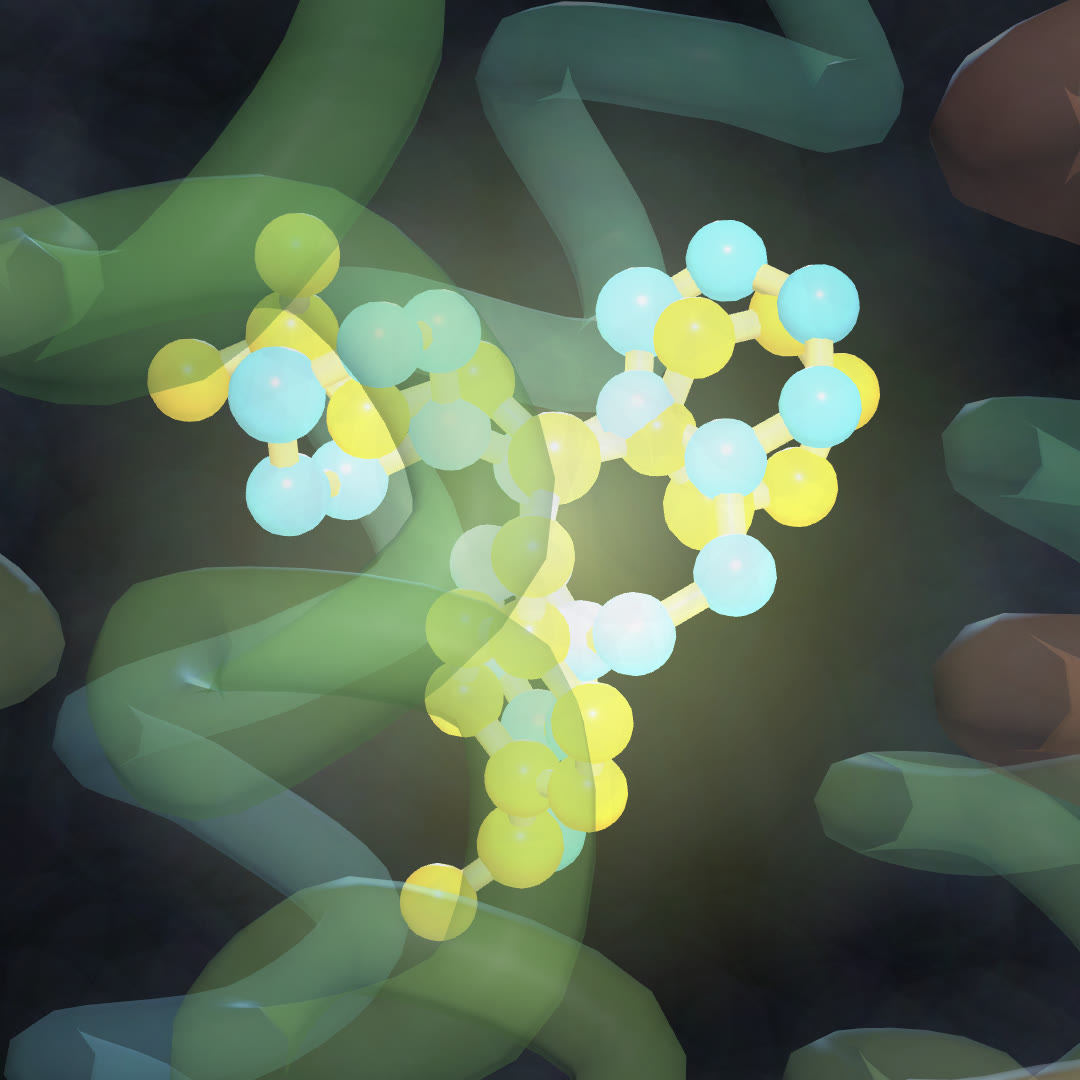 Two real cryo-EM structures of the human H1 receptor, overlaid: mepyramine, an older drowsy antihistamine (yellow, PDB 8X63), and desloratadine, a newer less-drowsy one (cyan, PDB 8X64).
In 2024 Wang et al. (Nat Commun) published cryo-EM structures of the human H1 receptor with several antihistamines bound, including mepyramine, a first-generation drug known for causing drowsiness, and desloratadine, a second-generation drug. We superimposed the two receptor structures on 263 matching backbone positions (root-mean-square deviation 1.13 Å) and moved both drugs into the same frame.
The two drugs sit in the same pocket. A cleaner test than eyeballing the overlay is to count, in each drug's own structure, the receptor amino acids within 4 Å of it. Desloratadine touches 9: Asp107, Tyr108, Ser111, Asn198, Trp428, Tyr431, Phe432, Phe435 and Tyr458. Mepyramine touches all 9 of those, plus five more (Ile115, Trp158, Thr194, Ala195, Ile454). At 4.5 Å the shared set is 13 of 13. (We first quoted the distance between the two drugs' centres, 0.59 Å, but that is smaller than the 1.13 Å error of the overlay itself, so it cannot be read that precisely.) At the receptor itself, the drowsy and the less-drowsy drug do the same job in the same place; the older one simply grips a little more.
A different door
If the lock is the same, the difference has to be somewhere else. The main one is getting into the brain. Chen et al. (Drug Metab Dispos 2003) compared mice with and without P-glycoprotein, a pump in the blood–brain barrier that pushes many drugs back out. For the three non-sedating antihistamines they tested, the brain-to-plasma ratio rose when the pump was missing: 2-fold for loratadine, 4-fold for cetirizine and more than 14-fold for desloratadine. For the sedating ones they tested (hydroxyzine, diphenhydramine and triprolidine), the ratio was about the same with or without the pump: they are not P-glycoprotein substrates. So the newer drugs are largely pumped back out of the brain, and the brain's own histamine signal keeps running.
That is not the whole story, and we changed one line of the video because of it. Our first draft said "the difference is the door, not the lock". Wang et al. point to two causes of the older drugs' side effects: crossing the blood–brain barrier, and poor receptor selectivity, meaning they also bind other receptors. Chen et al. did not test mepyramine itself. "The big difference is getting in" is as far as the evidence goes.
What this does not show
- The neurotransmitter labels in the fly connectome are predictions, not measurements of histamine in each cell.
- The eight gold neurons have not been shown to control sleep. The sleep evidence comes from whole-fly mutants.
- Fly and human histamine receptors are different proteins. Antihistamines designed for human H1 are not expected to act on fly receptors.
- The brain-entry evidence is from mice and cultured cells, not people.
- "Less-drowsy" is not "never drowsy". Second-generation drugs can still cause sleepiness in some people.
Methods and data
- Fly: Janelia FlyEM male CNS v1.0 (CC BY). Predicted transmitter labels (
consensusNt) and types from the public segment properties. Output synapses read per neuron from the public synapse table. Partners counted as distinct postsynaptic bodies, and as annotated neurons when the body appears in the release's neuron list. Visual system = superclassesol_sensoryandol_intrinsic. Code:verify/fly-histamine.js,data/fetch-fly-hist.js. - Structures: PDB 8X63 (mepyramine) and 8X64 (desloratadine), receptor chain B, superimposed by least squares on Cα atoms. Contact residues: any atom within 4 Å of the drug's heavy atoms. Code:
data/pdb-pair.js. - The figure of the fly renders real meshes. The light pulses in the video are illustrative.
References
- Berg S., Beckett I.R. et al. (2026). Sexual dimorphism in the complete Drosophila male central nervous system connectome. Cell 189:5504–5526. doi:10.1016/j.cell.2026.08.015
- Oh Y. et al. (2013). Histamine-HisCl1 receptor axis regulates wake-promoting signals in Drosophila melanogaster. PLoS One 8:e68269. doi:10.1371/journal.pone.0068269
- Haas H.L., Sergeeva O.A., Selbach O. (2008). Histamine in the nervous system. Physiol Rev 88:1183–1241. doi:10.1152/physrev.00043.2007
- Wang D. et al. (2024). Molecular mechanism of antihistamines recognition and regulation of the histamine H1 receptor. Nat Commun 15:84. doi:10.1038/s41467-023-44477-4
- Chen C. et al. (2003). P-glycoprotein limits the brain penetration of nonsedating but not sedating H1-antagonists. Drug Metab Dispos 31:312–318. doi:10.1124/dmd.31.3.312
More from Inside the Connectome: Human vs mouse vs fly: how we measured five rounds · Same lock, different effects: LSD and lisuride · Why is nicotine so hard to quit?
Author — the operator of kbrain-map.org, a researcher who analyses multi-omics (proteomics) data. The measurements behind the Inside the Connectome videos and this post were made directly on the public connectome and protein-structure data.
A note from the operator — The video is 46 seconds long, so it could not show everything behind the numbers. If you find a mistake, please tell us: we will fix the post and pin a correction under the video.
관련 글
Human vs Mouse vs Fly: How We Measured Five Rounds of Brain-Cell Comparisons (and What We Got Wrong First)
10월 16일 · 15 min read
NeuroscienceWhich H01 Is It? Two Public Synapse Releases, Only One Matches the Paper — and How We Found Out the Hard Way
10월 19일 · 8 min read
NeuroscienceWhy Is Nicotine So Hard to Quit? What the Real Receptor Structures Show
10월 3일 · 6 min read
NeuroscienceWe Followed One Neuron Through a Map of the Human Brain — Methods, Numbers, and What We Got Wrong
10월 1일 · 9 min read