Same Lock, Different Effects: Why LSD Is Psychedelic and Lisuride Isn't
LSD and lisuride occupy the same pocket of the human serotonin 2A receptor, 0.12 Å apart. Only LSD is psychedelic. We compared five published crystal structures residue by residue, checked the LSD pose against an independent structure from another lab, and went through what the literature says explains the difference.
한국어판: 같은 자물쇠, 다른 효과
This post started from our short What do LSD and magic mushrooms grab in your brain?. While making it, we overlaid LSD and lisuride in the serotonin 2A receptor and they landed almost exactly on top of each other. That raised a question a 40-second video can't answer.
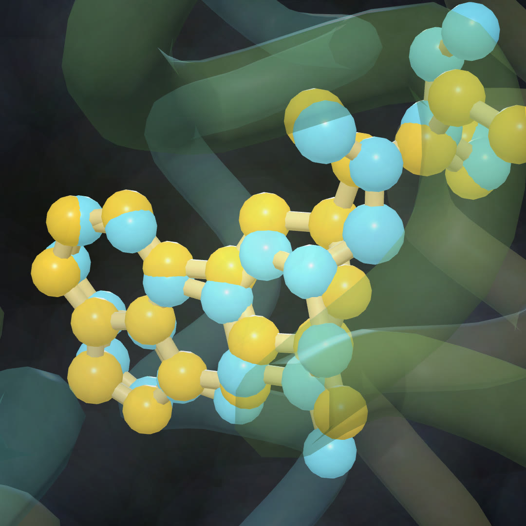 LSD (yellow, PDB 7WC6) and lisuride (cyan, PDB 7WC7) after superposing the two receptors. The ligand centers end up 0.12 Å apart.
The puzzle
Every classic psychedelic we know of, LSD, psilocybin and mescaline among them, is an agonist of the serotonin 2A receptor (5-HT2A) (González-Maeso et al. 2007). The receptor is not just a correlate. In healthy volunteers, the 5-HT2A antagonist ketanserin blocked the psychosis-like effects of psilocybin in a dose-dependent way (Vollenweider et al. 1998).
Lisuride complicates this. It is built on the same ergoline scaffold as LSD and it activates 5-HT2A, yet it lacks comparable psychoactive effects (González-Maeso et al. 2007). If the receptor is the switch, why does one of these near-identical keys flip it and the other doesn't?
What we looked at
In 2022, Cao and colleagues published crystal structures of the human 5-HT2A receptor bound to four ligands, all from the same construct and the same lab (Cao et al., Science 2022):
| PDB | Ligand | Resolution |
|---|---|---|
| 7WC4 | serotonin | 3.2 Å |
| 7WC5 | psilocin (the active form of psilocybin) | 3.2 Å |
| 7WC6 | LSD | 2.6 Å |
| 7WC7 | lisuride | 2.6 Å |
As an independent check we also used 6WGT, an LSD-bound 5-HT2A structure from a different group (Kim et al., Cell 2020). It has three copies of the receptor in one crystal. None of these five structures has a G protein attached, and in all of them a fusion protein sits in the third intracellular loop (residues numbered 1001 and up), which we excluded.
How we measured, briefly:
- Contacts: receptor residues with any heavy atom within 4.2 Å of the ligand, counted in each ligand's own structure, with the closest distance recorded.
- Superposition: least-squares fit on matched Cα atoms of the receptor chain.
- Asp155: the shortest distance from a ligand nitrogen to its carboxylate, since a salt bridge here is the classic anchor for aminergic ligands.
A mistake worth mentioning
Our first contact counts were taken after superposing everything onto the LSD structure. That gave lisuride 17 contacts. Recounting in lisuride's own crystal gave 19. The difference is small, but it shows how sensitive these counts are to the frame and the cutoff. Everything below uses each ligand's own structure.
The 6WGT check made the same point more strongly. Within that single crystal, the three copies of the LSD complex gave 14, 18 and 18 contacts. So we read the counts as rough measures of how much of the pocket a ligand fills, not as precise numbers. A difference like 17 against 19 means nothing.
Four footprints
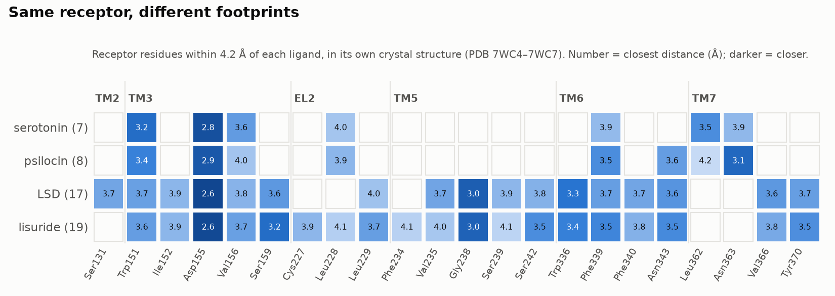
All four ligands make the Asp155 salt bridge (2.6 to 2.9 Å), and all four touch Trp151, Val156 and Phe339. That is the shared core of the binding site.
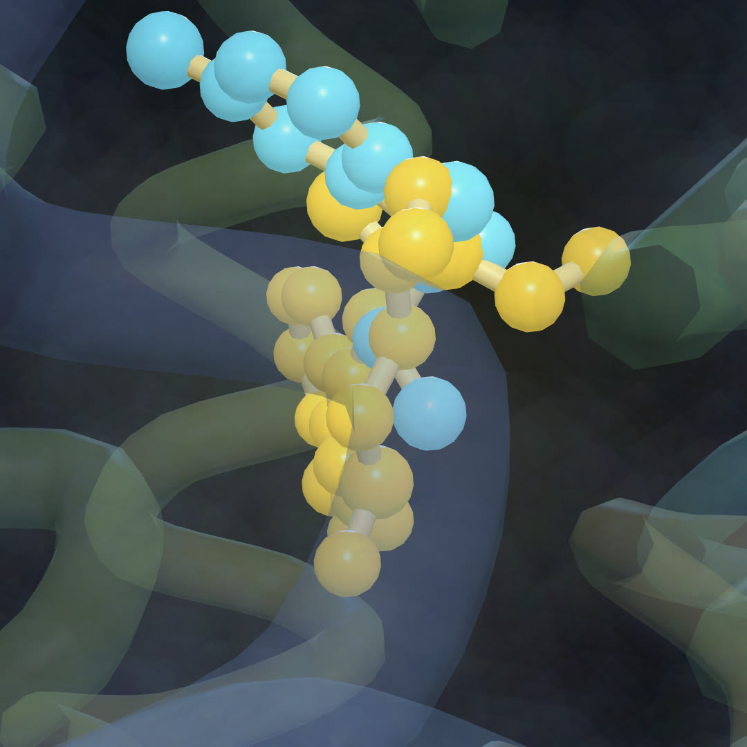
Beyond that core they diverge. In these crystals, serotonin and psilocin lean toward the seventh helix, touching Leu362 and Asn363. LSD and lisuride reach the other way, down between helices 5 and 6: Gly238, Ser242, Trp336, Phe340, Tyr370. Serotonin touches none of those here. The boundary is not sharp, though: one of the three LSD copies in 6WGT (below) also reaches Leu362 and Asn363.
| heavy atoms | contacts (4.2 Å) | N to Asp155 | |
|---|---|---|---|
| serotonin | 13 | 7 | 2.8 Å |
| psilocin | 15 | 8 | 2.9 Å |
| LSD | 24 | 17 | 2.6 Å |
| lisuride | 25 | 19 | 2.6 Å |
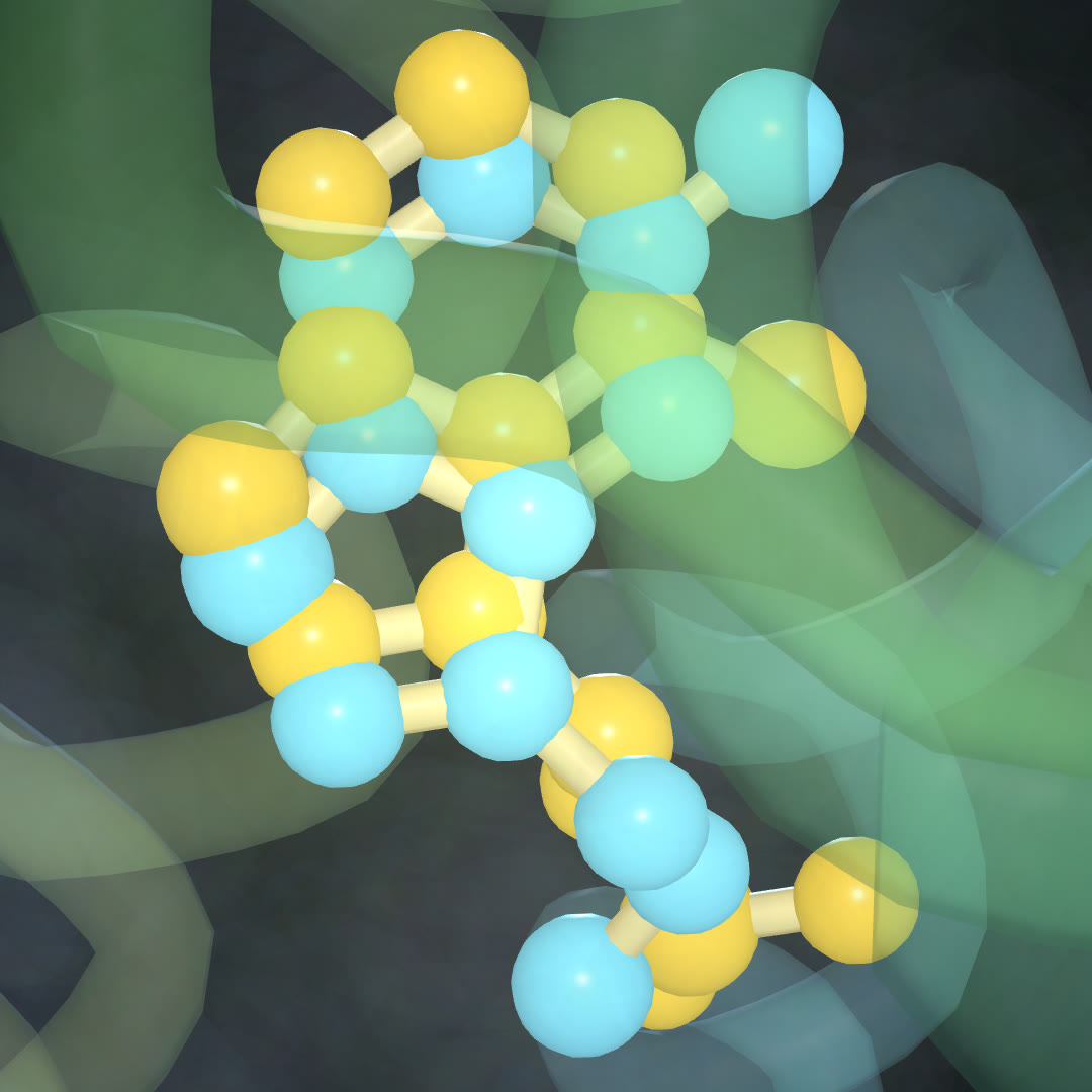
Psilocin is very nearly serotonin's double. Its center is 0.47 Å from serotonin's after superposition (receptor RMSD 0.37 Å), and it touches all seven of serotonin's contact residues plus Asn343.
| Psilocin vs serotonin | LSD vs serotonin |
|---|---|
 |  |
One thing we could not settle. Cao et al. report that serotonin and psilocin can take a second binding mode besides the canonical one. Their abstract doesn't say which mode these particular crystal poses represent, and the full text is not open access. So we describe the geometry and leave the question of which pose matters for signaling open.
Is the LSD pose real?
A single crystal structure can mislead, so we superposed all three LSD copies from 6WGT onto 7WC6. The LSD centers agree to within 0.5 to 0.7 Å. The deep contacts with Gly238, Ser242, Trp336 and Phe340 appear in every copy, from both labs. Atom by atom the ligand differs more, by 1.3 to 1.8 Å RMSD depending on the copy, so the fine details of the pose are less certain than its overall placement. The placement itself holds up.
LSD and lisuride, side by side
Superposing 7WC6 and 7WC7 gives a receptor RMSD of 0.38 Å. The ligand centers sit 0.12 Å apart, about a tenth of a carbon–carbon bond. Lisuride shares 16 of LSD's 17 contact residues and adds three at the extracellular end (Cys227, Leu228, Phe234). Both make the same 2.6 Å salt bridge with Asp155.
On this evidence, you could not tell from the binding pose which of the two is a psychedelic.
What explains it, then
A structure without a G protein shows where a drug sits, not what the receptor does next. Three lines of work go after the "what next".
Signaling pathways. In mouse cortex, González-Maeso et al. (2007) found that LSD and lisuride both drive the canonical 5-HT2A route through phospholipase C in the same neurons. LSD's response also involved pertussis-toxin-sensitive Gi/o proteins and Src, and lisuride's did not.
Binding kinetics. Wacker et al. found that LSD comes off both 5-HT2B and 5-HT2A exceptionally slowly. Their simulations point to a "lid" formed by extracellular loop 2. A mutation expected to loosen that lid sped up LSD's binding kinetics and selectively weakened its β-arrestin2 recruitment (Wacker et al., Cell 2017).
How hard the receptor is pushed. The most direct answer so far comes from Wallach et al., who built a series of 5-HT2A-selective agonists with graded Gq efficacy (Wallach et al., Nat Commun 2023). In male mice, Gq efficacy predicted the head-twitch response, a standard proxy for psychedelic effects. β-arrestin2 recruitment did not. Below a threshold of Gq activation there was no head twitch, and the non-psychedelic analogs fell under about 70% of serotonin's maximum. The paper cites a calcium-mobilization Emax of 48.6% for lisuride against 84.6% for LSD.
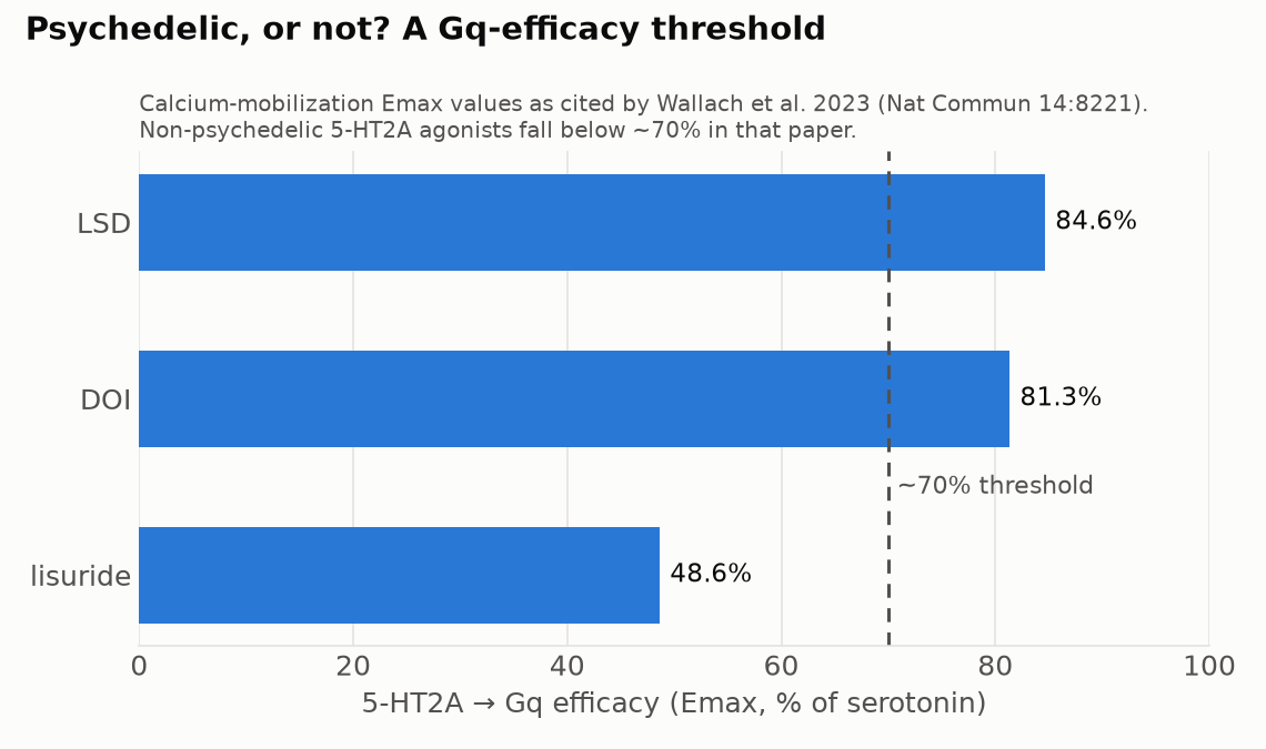
That fits the Cao et al. result too: their β-arrestin-biased 5-HT2A agonists had antidepressant-like effects in mice without hallucinogen-like behavior.
One wrinkle. Kim et al. (2020) describe LSD itself as an "arrestin-biased" ligand, yet in Wallach's assays LSD has strong Gq efficacy. "Bias" is always relative to a reference ligand and to particular assays, and these papers do not use the same ones. Comparing bias labels across studies should be done with care.
Our working summary is: same seat, different push. LSD and lisuride sit in essentially the same place. They differ in how strongly they drive the receptor toward Gq signaling, and possibly in how long they stay bound. Most of that difference is not visible in a static crystal structure. The field has not settled on a single mechanism, and the head-twitch threshold is a mouse measure, not a human one.
For another receptor read the same way, structure by structure, see Why Is Nicotine So Hard to Quit? What the Real Receptor Structures Show.
Limits
- The five structures are static, have no G protein, and range from 2.6 to 3.4 Å resolution. They cannot show efficacy.
- Contact counts depend on the cutoff and on which copy you use (14 to 18 across the three 6WGT copies). Small differences in counts are not meaningful.
- Emax values depend on the assay. The numbers above are the ones Wallach et al. cite.
- Lisuride also acts at other receptors, including 5-HT1A, which shapes its overall effects.
References
- Cao D. et al. (2022). Structure-based discovery of nonhallucinogenic psychedelic analogs. Science 375. doi:10.1126/science.abl8615 (PDB 7WC4–7WC7)
- Kim K. et al. (2020). Structure of a hallucinogen-activated Gq-coupled 5-HT2A serotonin receptor. Cell. doi:10.1016/j.cell.2020.08.024 (PDB 6WGT)
- Wallach J. et al. (2023). Identification of 5-HT2A receptor signaling pathways associated with psychedelic potential. Nat Commun 14: 8221. doi:10.1038/s41467-023-44016-1
- Wacker D. et al. (2017). Crystal structure of an LSD-bound human serotonin receptor. Cell 168: 377–389. doi:10.1016/j.cell.2016.12.033
- González-Maeso J. et al. (2007). Hallucinogens recruit specific cortical 5-HT2A receptor-mediated signaling pathways to affect behavior. Neuron. doi:10.1016/j.neuron.2007.01.008
- Vollenweider F.X. et al. (1998). Psilocybin induces schizophrenia-like psychosis in humans via a serotonin-2 agonist action. NeuroReport. doi:10.1097/00001756-199812010-00024
Structures from the RCSB Protein Data Bank (CC0). Contacts, superpositions and renders are our own analysis of the public files; see our Methodology page. This is a post about brain science. It is not medical advice and not an endorsement of drug use.
관련 글
Why Is Nicotine So Hard to Quit? What the Real Receptor Structures Show
10월 3일 · 6 min read
신경과학같은 자물쇠, 다른 효과 — LSD는 환각을 일으키고 리수라이드는 왜 아닐까
10월 9일 · 15 min read
NeuroscienceWe Followed One Neuron Through a Map of the Human Brain — Methods, Numbers, and What We Got Wrong
10월 1일 · 8 min read
NeuroscienceDoes Chronic Stress Really Lower Hippocampal BDNF? What an LLM Read of 9,585 Studies Actually Says
6월 5일 · 8 min read