Does Parkinson's Start Only After 80% of Neurons Die? We Traced the Number Back to Its Sources
A widely repeated line says 80% of dopamine neurons are already gone when Parkinson's symptoms appear. We read the studies behind it. For the cells themselves, the best estimate is closer to 30%; the 80% is about something else.
This is the companion post to our Short Parkinson's symptoms only after 80% of neurons die? Not quite. The video is under a minute long. This post has the numbers and where each came from.
Educational content, not medical advice. If you have questions about Parkinson's disease or your own health, please talk to a doctor.
한국어판: 파킨슨병은 뉴런이 80% 죽어야 시작된다? 그 숫자의 출처를 따라가 봤습니다
The line you have probably heard
"By the time Parkinson's symptoms appear, 80% of the brain's dopamine neurons are already dead."
It turns up in lectures, news stories and patient leaflets. It is easy to remember, and it carries a grim message: by the time anyone notices, almost everything is lost.
We wanted to know where the number came from, so we went back to the studies behind it and read what they actually measured.
The short answer
For the cells themselves, the best estimate is closer to 30%. A 2010 review of the studies of brains donated after death concluded that movement symptoms appear when there is "about a 30% loss" of these neurons, compared with people of the same age (Cheng et al., Annals of Neurology 2010). The neurons live in the substantia nigra, a small region in the midbrain.
The 80% is about something else: how much dopamine is left in the striatum, the region where those neurons send their branches and release dopamine. One older estimate of that loss at the onset of symptoms is around 70–80%, but the study most often given as the source cannot support the number.
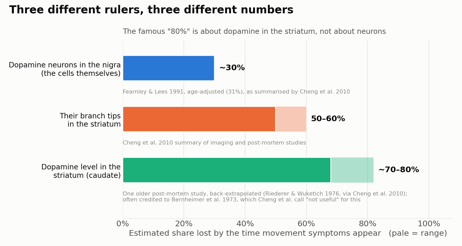 Figure 1. "How much is lost when symptoms appear?" has three different answers, depending on what you measure. The pale part of a bar is the range of estimates. Sources: cell bodies, Fearnley & Lees 1991 (age-adjusted), as summarised by Cheng et al. 2010; branch tips, Cheng et al.'s summary of imaging and post-mortem studies; striatal dopamine, one older post-mortem study of the caudate back-extrapolated to onset (Riederer & Wuketich 1976, as analysed by Cheng et al.) Chart: ours, drawn from the numbers in those papers.
Where the 80% came from
Dopamine neurons have their cell bodies in the substantia nigra, but their long branches end far away, in the striatum. That is where they release dopamine.
In 1973, Herbert Bernheimer and colleagues measured dopamine in the brains of people who had died with several kinds of parkinsonism (Bernheimer et al., Journal of the Neurological Sciences 1973). They reported that even mild, recently noticeable parkinsonism went with a large shortfall of dopamine in the striatum. This study is often cited for the claim that symptoms begin when about 80% of striatal dopamine is gone.
When Cheng and colleagues reviewed the evidence, they were direct about it: the Bernheimer study "is often cited in support of the statement that Parkinson motor signs first appear when about 80% of striatal or putaminal dopamine is lost … However, the Bernheimer study does not provide useful information for this estimate." Only 13 people with Parkinson's disease had their dopamine measured, and the study did not estimate the level at onset directly. One older post-mortem study of the caudate (part of the striatum), back-extrapolated to onset, gives 68% and 82% in two groups of patients (Riederer & Wuketich 1976, as analysed by Cheng and colleagues), so the order of magnitude for dopamine is not unreasonable.
What does not follow is the version that gets repeated most: 80% of neurons. That figure is about the chemical in the striatum, not about how many cells in the nigra have died.
Where the 30% comes from
In 1991, Jeremy Fearnley and Andrew Lees counted the dopamine neurons in donated brains from people with and without Parkinson's disease (Fearnley & Lees, Brain 1991). Most of these neurons contain a dark pigment, neuromelanin, which makes them easy to count.
Working back from how long each person had been ill, their own abstract puts the loss at the time symptoms begin at 48% for the part of the nigra they counted, and 68% in its hardest-hit tier. But some of these cells are lost with normal ageing anyway. Adjusted for age, the same regression gives about 31%, as Cheng and colleagues report. Two later studies point the same way: for one, Cheng and colleagues extrapolated the figure themselves (Ma et al. 1997); for the other, they report a 30% estimate (Greffard et al. 2006). Their summary: "at the time of first diagnosis of PD, only 30% or so of dopamine neurons of the SN have been lost and only 50-60% of their axon terminals." (Most textbooks and reviews say about 50%, Cheng and colleagues note; the age adjustment is why their estimate is lower.)
Two honest caveats. The estimate comes from a few brains and, in Fearnley and Lees's case, a single slice of the nigra. "About 30%" is the best-argued figure, not a precise constant.
Many neurons stop making dopamine before they die
There is a second reason the numbers disagree. "Losing a dopamine neuron" can mean two things: the cell has died, or the cell is still there but no longer makes dopamine.
In 2013, Jeffrey Kordower and colleagues counted both in the same donated brains, from people who had died at different times after their diagnosis (Kordower et al., Brain 2013). One count used the enzyme a neuron needs to make dopamine (tyrosine hydroxylase, TH). The other used the pigment, which stays in the cell either way.
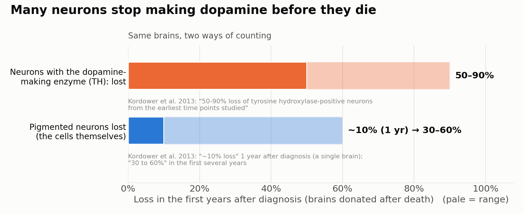 Figure 2. In the same brains, neurons with the dopamine-making enzyme disappear earlier and more widely than the pigmented cells do. The enzyme count includes cells that have died. Source: Kordower et al. 2013 ("50-90% loss of tyrosine hydroxylase-positive neurons from the earliest time points studied"; "only a ∼10% loss of melanized neurons in the one case evaluated 1 year post-diagnosis, and variable (30 to 60%) loss during the first several years"). The 10% comes from a single brain. Chart: ours.
Counted by the enzyme, the loss looks severe from early on. Counted by the cells themselves, many are still there. Both are true; they answer different questions. The same study also found that the dopamine markers of the branch tips in the most affected part of the striatum (the dorsal putamen) were almost completely gone within about four years of diagnosis.
Ten years of ageing, ten years of disease
Everyone loses some of these neurons with age. Fearnley and Lees measured how fast, in the same part of the nigra.
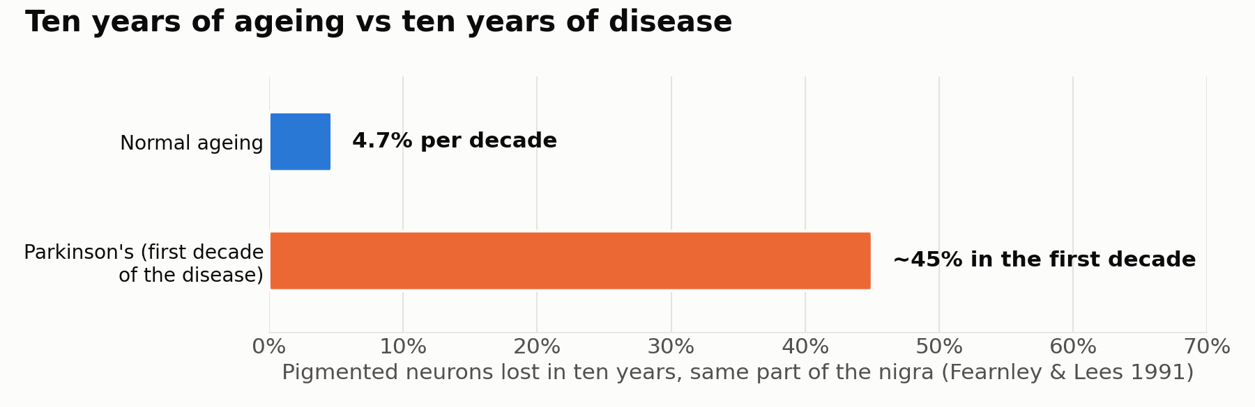 Figure 3. Pigmented neurons lost over ten years in the same part of the nigra: about 4.7% per decade with normal ageing, about 45% in the first decade of Parkinson's disease (20 cases). Source: Fearnley & Lees 1991. Chart: ours.
The difference is what makes Parkinson's a disease rather than ordinary ageing: in the first decade of the disease, the same cells are lost roughly ten times faster.
What a brain scan actually sees
If cells can stay alive but stop working, what do brain scans measure?
One scan sometimes used to support a clinical diagnosis, DaTscan, does not look at the cell bodies at all. It uses a small amount of a radioactive tracer, ioflupane, that attaches to the dopamine transporter (DAT): a protein on the branch tips that pulls dopamine back in after it has been released (Bajaj et al., J Neurol Neurosurg Psychiatry 2013). Fewer working branch tips in the striatum means a weaker signal. So the scan is a stand-in for the middle bar in Figure 1, not the first one. (Doctors diagnose Parkinson's mainly from symptoms and examination; the scan can also be abnormal in some related conditions.)
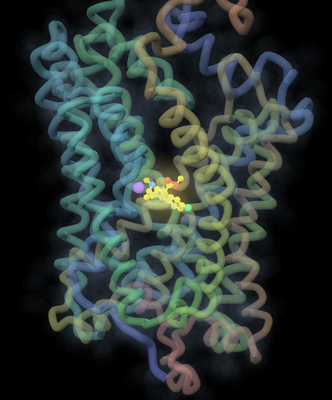 Figure 4. The dopamine transporter, the protein the DaTscan tracer attaches to, with a related test molecule (yellow) sitting in its central pocket. Technical details are in "How we checked" below. Render: ours, from public data.
What this means, and what it does not
In plain terms: when Parkinson's is diagnosed, most of these neurons are typically still alive, and in the first years after diagnosis many of them no longer show the dopamine-making enzyme. Their branch tips are hit harder and faster. "80% is already gone" and "about 30% of the cells are gone, and many of the rest are struggling" leave very different impressions. The second is one reason researchers work on detecting the disease earlier and on ways to protect the remaining cells.
So far, no treatment has been proven to slow the disease itself. Whether struggling neurons can be protected or rescued is still being researched. We are not saying anything here about treatment, outlook or what any individual should do; those are questions for doctors and clinical research.
How to read a "how much is lost" number
Next time you meet a percentage like this, three questions sort out most of the confusion:
- What was counted? The cells, their branch tips, or the amount of dopamine?
- When? At symptom onset, at diagnosis, or at death?
- How? By pigment (the cell is there) or by an enzyme (the cell is still making dopamine)?
The 30% and the 80% are not two guesses at the same thing. They are answers to different questions.
How we checked
- We read the abstracts and, where open, the full texts of the studies cited here. Every number in the figures is quoted from those papers. The charts are drawn by a script from a single table (
blog/parkinsons-80/figs.pyin theshoo99/science-shortsrepository), so no number is retyped by hand. - "About 30%" is the conclusion of the Cheng et al. 2010 review. The 31% comes from Fearnley & Lees's age-adjusted regression, as Cheng et al. report it; Fearnley & Lees's own abstract gives 48% (caudal nigra) and 68% (its hardest-hit tier) without that adjustment. Ma's ~30% is Cheng et al.'s extrapolation; Greffard's 30% is as reported by Cheng et al. (it is not in Greffard's abstract).
- The 70–80% striatal dopamine range is one older post-mortem study of the caudate, back-extrapolated to onset (68% and 82% in two patient groups; Riederer & Wuketich 1976, as analysed by Cheng et al.). Cheng et al. note that post-mortem dopamine measurements are affected by the delay between death and measurement. Imaging studies with transporter tracers give 50–70% loss in the putamen at onset (also cited by Cheng et al.).
- Bernheimer et al. (1973) studied several kinds of parkinsonism; only 13 people with Parkinson's disease had dopamine measured. The step from "80% of striatal dopamine" to "80% of neurons" is common in popular retellings; we found no study that measured 80% of nigral neurons lost at onset.
- Figure 4 is our render of PDB entry 8VBY: an experimental cryo-electron-microscopy structure of a stabilised human dopamine transporter with β-CFT (yellow), a cocaine analogue from the same chemical family as ioflupane (phenyltropanes), at 3.19 Å resolution. The purple sphere is a sodium ion. The deposited model also contains a second inhibitor and a zinc ion, not shown. No structure of the transporter with ioflupane itself was available to us (Srivastava et al., Nature 2024).
- We could not open the full text of one older counting study (Pakkenberg et al. 1991, a scanned PDF), so we used only its abstract.
- Before publishing, an adversarial fact-check pass tried to break each number and sentence; it caught that our first draft credited the 31% to the wrong authors, placed the two bars in Figure 3 in different regions when they are the same, and called the 80% a pure invention when the dopamine estimate has some support. A second pass read the post as a non-specialist and marked confusing terms and figures; we simplified both.
References
- Cheng H.C., Ulane C.M., Burke R.E. (2010). Clinical progression in Parkinson disease and the neurobiology of axons. Ann Neurol 67:715–725. doi:10.1002/ana.21995
- Fearnley J.M., Lees A.J. (1991). Ageing and Parkinson's disease: substantia nigra regional selectivity. Brain 114:2283–2301. doi:10.1093/brain/114.5.2283
- Bernheimer H. et al. (1973). Brain dopamine and the syndromes of Parkinson and Huntington. Clinical, morphological and neurochemical correlations. J Neurol Sci 20:415–455. doi:10.1016/0022-510X(73)90175-5
- Riederer P., Wuketich S. (1976). Time course of nigrostriatal degeneration in Parkinson's disease. J Neural Transm 38:277–301. doi:10.1007/BF01249445
- Kordower J.H. et al. (2013). Disease duration and the integrity of the nigrostriatal system in Parkinson's disease. Brain 136:2419–2431. doi:10.1093/brain/awt192
- Ma S.Y. et al. (1997). Correlation between neuromorphometry in the substantia nigra and clinical features in Parkinson's disease using disector counts. J Neurol Sci 151:83–87. doi:10.1016/S0022-510X(97)00100-7
- Greffard S. et al. (2006). Motor score of the Unified Parkinson Disease Rating Scale as a good predictor of Lewy body–associated neuronal loss in the substantia nigra. Arch Neurol 63:584–588. doi:10.1001/archneur.63.4.584
- Pakkenberg B. et al. (1991). The absolute number of nerve cells in substantia nigra in normal subjects and in patients with Parkinson's disease estimated with an unbiased stereological method. J Neurol Neurosurg Psychiatry 54:30–33. doi:10.1136/jnnp.54.1.30
- Bajaj N., Hauser R.A., Grachev I.D. (2013). Clinical utility of dopamine transporter single photon emission CT (DaT-SPECT) with (123I) ioflupane in diagnosis of parkinsonian syndromes. J Neurol Neurosurg Psychiatry 84:1288–1295. doi:10.1136/jnnp-2012-304436
- Srivastava D.K. et al. (2024). Structure of the human dopamine transporter and mechanisms of inhibition. Nature 632:672–677. doi:10.1038/s41586-024-07739-9
More from Inside the Connectome: Is dopamine the pleasure chemical? Not quite · Why do some allergy pills make you sleepy?
Author — the operator of kbrain-map.org, a researcher who analyses multi-omics (proteomics) data. The charts were drawn from the numbers in the cited papers; the structure image is our own render of public data.
A note from the operator — This post is about where a number comes from, not about the care of any person. If you find a mistake, please tell us and we will correct the post.
Educational content, not medical advice.
Related
Is Dopamine the Pleasure Chemical? Not Quite. 390 Fly Neurons, One Receptor, and What the Experiments Say
Oct 27 · 13 min read
NeuroscienceWhy Do Some Allergy Pills Make You Sleepy? Eight Histamine Neurons, One Receptor Pocket, and a Door
Oct 20 · 11 min read
NeuroscienceWhy Is Nicotine So Hard to Quit? What the Real Receptor Structures Show
Oct 3 · 6 min read
NeuroscienceNeuroscience of Addiction Explained — How the Brain Gets Hijacked
Feb 20 · 10 min read