What Is in Layer 1 of the Human Cortex? Few Neurons, Many Astrocytes
In a cubic millimetre of human cortex (H01), only 18% of layer-1 cells are neurons (827 of 4,595), against 70% in layer 2. The most common cell is the astrocyte: 37%. What else is there, why the layer is mostly wiring, and the caveats.
This is the companion post to two of our Shorts: Why does the top layer of your brain have so few neurons? and The top layer of your cortex is full of star cells. Both numbers are on our Numbers page: few neurons and astrocytes.
The short answer
Layer 1 is the outermost layer of the cortex, just under the brain's surface. In one sample of human cortex we counted 4,595 cell bodies labelled as layer 1. Only 827 of them (18%) are neurons. One layer down, in layer 2, neurons are 70% of the cells (4,656 of 6,651).
The most common cell in layer 1 is not a neuron. It is the astrocyte, a star-shaped support cell: 1,688 cells, 37%.
Layer 1 is mostly wiring. It is where the branch tips of neurons from deeper layers meet signals arriving from other parts of the brain.
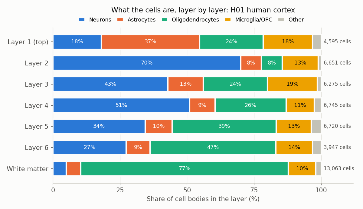
What each layer's cell bodies are labelled as. "Other" = cells marked unknown, blood-vessel cells and C-shaped glia. Our count from H01 somas.csv, using the dataset's own cell-type and layer labels.
The full count for layer 1
| Cell type | Cells in layer 1 | Share |
|---|---|---|
| Astrocytes | 1,688 | 36.7% |
| Oligodendrocytes | 1,086 | 23.6% |
| Microglia / OPC | 845 | 18.4% |
| Neurons | 827 | 18.0% |
| Unknown | 121 | 2.6% |
| Blood-vessel cells | 28 | 0.6% |
| Total | 4,595 |
The 827 neurons are mostly inhibitory interneurons: 586 (71%). The rest are 156 unclassified neurons, 72 pyramidal cells and 13 spiny atypical cells.
The map we used
H01 is a fragment of human temporal cortex removed during epilepsy surgery, imaged with an electron microscope and reconstructed in 3D (Shapson-Coe et al. 2024, Science). It is about one cubic millimetre, cut as a slab that runs through all six cortical layers into the white matter below. It is public under CC BY 4.0.
Its somas.csv table lists 49,379 cell bodies, each with a position, a cell type (classified by hand by the H01 team) and a layer. We simply counted rows by layer and type. 1,383 cells have no layer label and are left out of the layer counts.
Why so few neurons?
Most of the cortex's neurons have their cell bodies in layers 2 to 6. Layer 1 holds their far ends instead. Pyramidal neurons send a long dendrite up toward the surface, and it branches into a "tuft" in layer 1. A review by Schuman et al. (2021, Annual Review of Neuroscience) describes layer 1 as "the predominant input layer for top-down information, relayed by a rich, dense mesh of long-range projections that provide signals to the tuft branches" of pyramidal cells.
The H01 paper's own measurement of what fills each layer agrees (Table S4, share of the segmented volume):
- In layer 1, cell bodies take up only 2.1% of the volume. Axons take 41.4% and dendrites 31.7%.
- In layers 2 to 6, cell bodies take up 9.6% to 12.9%.
So a cube of layer 1 is mostly cables, with relatively few cell bodies between them. The neurons that do live there are almost all interneurons in our count: 586 of the 671 neurons with an excitatory or inhibitory label (87%). We cover that in What fraction of neurons are inhibitory in the human cortex?.
Why so many astrocytes?
Astrocytes are glial cells: brain cells that are not neurons. In this sample, layer 1 is crowded with them:
- 1,688 of the sample's 5,474 astrocytes (30.8%) sit in layer 1, a thin layer near the surface.
- In every other layer, astrocytes are only 5% to 13% of the cells.
- The paper's density table (Table S6, corrected for tissue compression) gives 12,210 astrocytes per mm³ in layer 1, against 3,592 to 4,743 in layers 2 to 6.
- By volume, astrocytes fill 21.6% of layer 1, against 14.3% to 15.3% in layers 2 to 6 (Table S4).
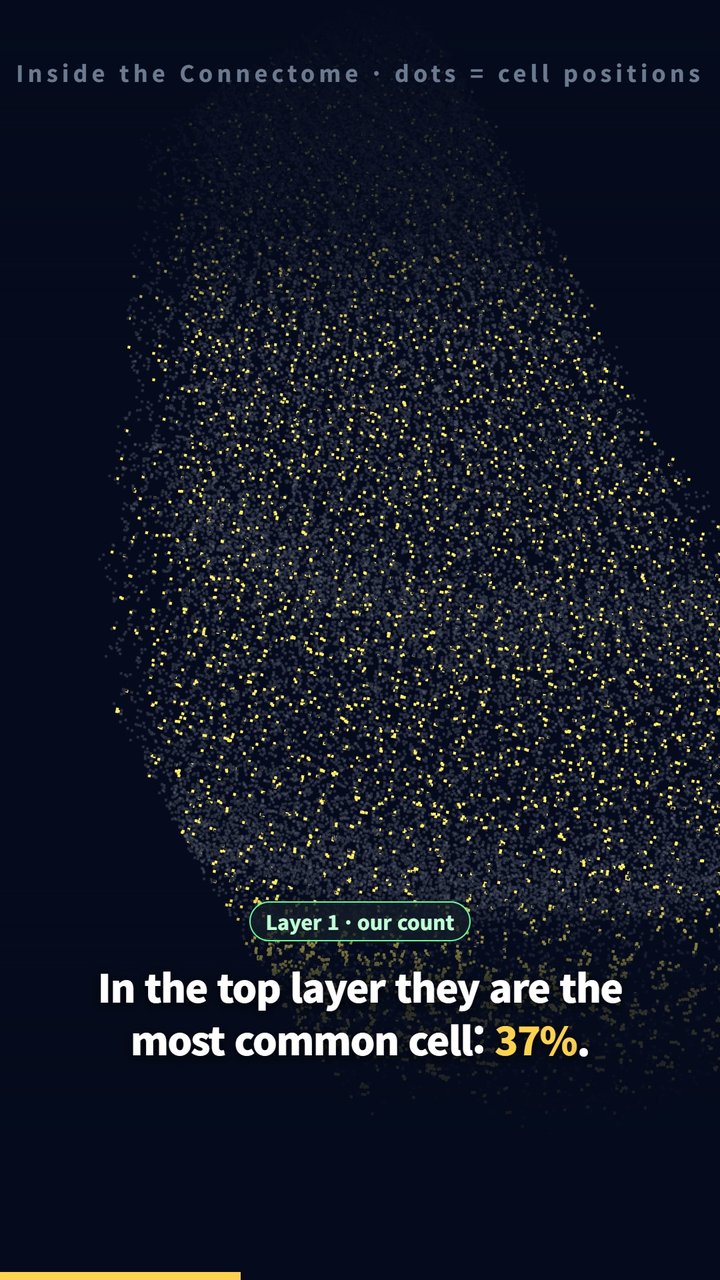
Frame from our Short. Gold dots: astrocytes. Grey dots: all other cells. Dots mark cell-body positions, not cell shapes.
Human astrocytes are also different from rodent ones. Oberheim et al. (2009, J Neurosci) found that human protoplasmic astrocytes are 2.6-fold larger in diameter than their rodent counterparts, and that human neocortex "harbors several anatomically defined subclasses of astrocytes not represented in rodents". One of them, the interlaminar astrocyte, "abundantly populates the superficial cortical layers and extends long processes" down to layers 3 and 4.
Our map does not tell astrocyte subtypes apart. We cannot say which of the 1,688 layer-1 astrocytes are interlaminar, only that layer 1 has far more astrocytes than any other layer.
What this does not tell you
- One sample. One person, one area of temporal cortex, removed during epilepsy surgery. Whether it is typical of healthy cortex is an open question the H01 authors raise themselves.
- Labels are the dataset's. Layer borders were drawn by the H01 team from cell positions and sizes. A cell near a border could be counted in either layer. The paper's density table lists no spiny (excitatory) neurons in layer 1, while our count from layer labels finds 72 pyramidal cells there; we have not checked where those cells sit.
- Counts are not densities. The table above counts cells per layer. Layers differ in thickness, so for a fair comparison between layers use the paper's per-mm³ values (we quote some above).
- Cell bodies only. Neurons with cell bodies in deeper layers still reach into layer 1 through their dendrites and axons. "Few neurons in layer 1" means few neuron cell bodies, not little neural activity.
Code and data
- H01
somas.csv: columnscelltypeandlayer; counts of rows per layer and type. - Volume shares and densities: the H01 paper's supplementary Tables S4 and S6.
- The chart comes from one re-runnable Python script.
Related
- How close are neurons to blood vessels?: the same
somas.csv, measured against the vessel network. - Which brain cells spread out evenly?: microglia and OPCs, the one class that does not change with layer.
References
- Shapson-Coe A, Januszewski M, Berger DR, et al. A petavoxel fragment of human cerebral cortex reconstructed at nanoscale resolution. Science. 2024;384:eadk4858. doi:10.1126/science.adk4858
- Schuman B, Dellal S, Prönneke A, Machold R, Rudy B. Neocortical layer 1: an elegant solution to top-down and bottom-up integration. Annu Rev Neurosci. 2021;44:221–252. doi:10.1146/annurev-neuro-100520-012117
- Oberheim NA, Takano T, Han X, et al. Uniquely hominid features of adult human astrocytes. J Neurosci. 2009;29:3276–3287. doi:10.1523/JNEUROSCI.4707-08.2009
Related
What Fraction of Neurons Are Inhibitory in the Human Cortex? About 31% in Our Count (vs 12% in Mouse)
Nov 2 · 6 min read
NeuroscienceWhat Is the Most Common Cell in the Human Brain? In This Sample, Not the Neuron
Nov 2 · 6 min read
NeuroscienceWhy Is White Matter White? 31.5% of Its Mapped Volume Is Myelinated Axons
Nov 2 · 7 min read
NeuroscienceHow Close Are Neurons to Blood Vessels? We Measured 10,441 of Them in a Real Human Brain Sample
Oct 30 · 6 min read