Why Is Nicotine So Hard to Quit? What the Real Receptor Structures Show
We overlaid two published cryo-EM structures of the human α4β2 nicotinic receptor, one holding nicotine and one holding acetylcholine. In all three pockets the two molecules sit about 1 Å apart. Here is what that shows, and what it doesn't.
Companion post to our short: Why is nicotine so hard to quit?. The short is 43 seconds; this post has the numbers, the method, and the caveats.
한국어판: 니코틴은 왜 끊기 어려울까
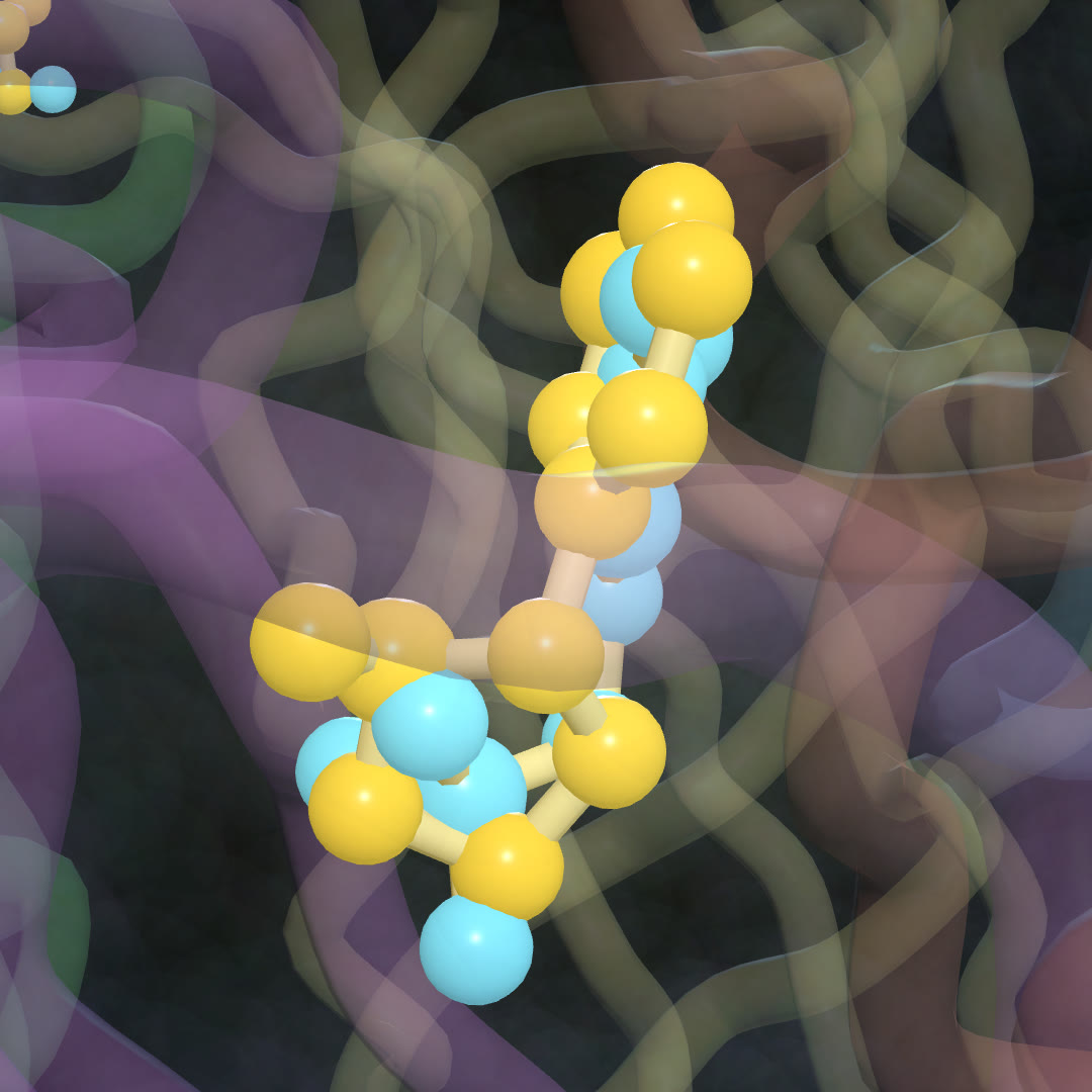 Nicotine (yellow, PDB 6CNK) and the brain's own messenger acetylcholine (cyan, PDB 8ST2), superposed in the same binding pocket of the human α4β2 receptor. Rendered from public PDB data (CC0).
The lock: the α4β2 nicotinic receptor
Nicotine acts on nicotinic acetylcholine receptors, ion channels that open when acetylcholine binds. The α4β2 type is the most abundant nicotinic receptor in the human brain and the principal target in nicotine addiction (Walsh et al., Nature 2018).
It is built from five subunits arranged around a central pore. α4β2 comes in two assemblies, 2α:3β and 3α:2β. The structures below are the 3α:2β form, which has three binding pockets: two at α4–β2 interfaces and one at an α4–α4 interface.
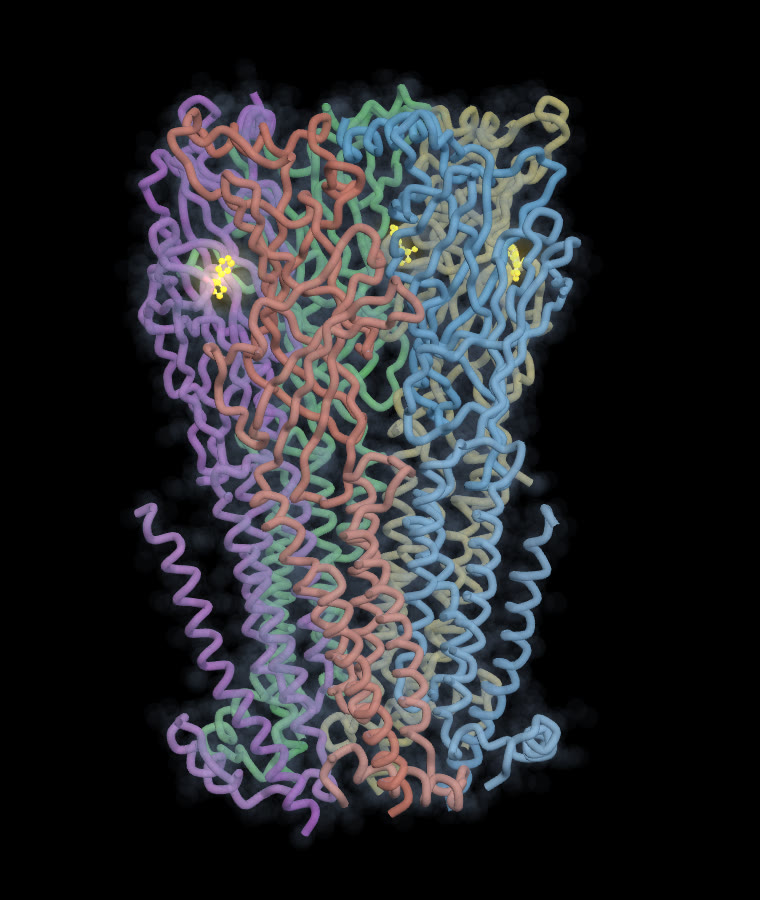 The whole receptor (five chains, colored separately). The small yellow molecules near the top are nicotine in its pockets. The lower half crosses the cell membrane.
The data
| Structure | What's bound | Method | Resolution | Source |
|---|---|---|---|---|
| 6CNK | nicotine (3 molecules) | cryo-EM | 3.9 Å | Walsh et al., Nature 2018 |
| 8ST2 | acetylcholine (3 molecules) | cryo-EM | 2.94 Å | Br J Pharmacol 2024, doi:10.1111/bph.16321 |
Both are the same human 3α:2β assembly with the same chain names (α4 = A, B, D; β2 = C, E). Both also contain mouse antibody fragments used to help the imaging; we left those out of the pictures.
Method
- We matched 1,822 Cα atoms between the two structures by chain and residue number (chains A–E).
- We superposed 8ST2 onto 6CNK by least squares (quaternion method). Overall RMSD 1.14 Å, so the two receptors are essentially the same shape.
- We moved acetylcholine into the 6CNK frame and measured how far its center sits from nicotine's center in each pocket.
Finding 1 — Same pocket, same size
| Pocket (ligand chain) | Interface | Nicotine ↔ acetylcholine centers |
|---|---|---|
| B | α4–β2 | 1.05 Å |
| D | α4–β2 | 1.31 Å |
| A | α4–α4 | 1.30 Å |
In every pocket, the two molecules sit about one ångström apart, less than the length of a single chemical bond. Nicotine is not "near" the acetylcholine site. It is the acetylcholine site.
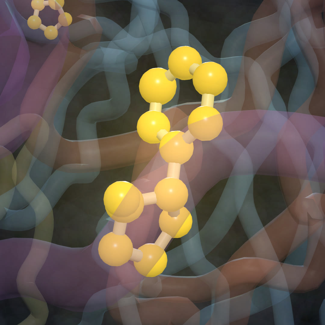
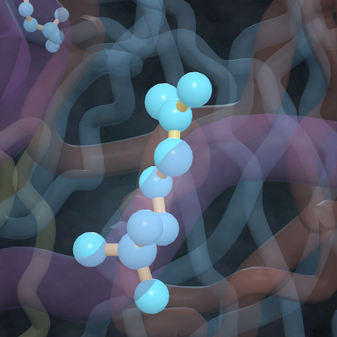
They are also the same size: 26 atoms each including hydrogens (nicotine C₁₀H₁₄N₂, acetylcholine C₇H₁₆NO₂⁺). Without hydrogens, nicotine has 12 heavy atoms and acetylcholine 10.
| Nicotine alone | Acetylcholine alone |
|---|---|
 |  |
Finding 2 — A charged nitrogen over a tryptophan ring
Why does nicotine grip brain receptors so tightly? Xiu et al. showed that at α4β2, nicotine's positively charged nitrogen makes a strong cation–π interaction with a tryptophan ring (TrpB). At muscle receptors this interaction is much weaker, even though the same tryptophan is there (Xiu et al., Nature 2009).
In 6CNK we measured the distance from nicotine's charged pyrrolidine nitrogen to the center of the Trp156 ring:
| Interface | N⁺ ↔ Trp ring | |
|---|---|---|
| B | α4–β2 | 4.1 Å |
| D | α4–β2 | 4.9 Å |
| A | α4–α4 | 5.4 Å |
These are within the range for cation–π contact at the α4–β2 sites. Caveat: at 3.9 Å resolution, side-chain positions carry real uncertainty, so read these as "in contact", not as precise values.
Finding 3 — The snapshot shows a tired receptor
Nicotine is an agonist: like acetylcholine, it opens the channel. That is what the short means by "it turns the key."
But the 6CNK snapshot is not an open channel. The authors describe it as a "putative closed-desensitized conformation": the receptor after prolonged exposure, shut and unresponsive (Walsh et al. 2018).
That links to the rest of the story:
- Acetylcholine is cleared fast. Acetylcholinesterase breaks it down within milliseconds at the synapse. Nicotine is not broken down there and stays much longer (textbook pharmacology).
- Receptors wear out. With nicotine lingering, α4β2 receptors spend more time in exactly this desensitized state.
- The brain makes up for it. In post-mortem brains of smokers, nicotinic binding sites were 250–400% of nonsmokers' levels in the cortex (Perry et al., J Pharmacol Exp Ther 1999).
- This receptor drives the reward. Mice lacking the β2 subunit showed no nicotine-evoked dopamine release in the ventral striatum, and self-administered less nicotine (Picciotto et al., Nature 1998).
What the short simplified
| In the short | More precisely |
|---|---|
| "It turns the key, and the channel opens" | Nicotine is an agonist and opens the channel. The structure shown, however, is the closed-desensitized state that follows. |
| "Receptors wear out" | Desensitization: prolonged agonist exposure leaves the receptor bound but closed. |
| "The brain builds more of them" | Upregulation of nicotinic binding sites, measured in smokers' brains (Perry 1999). The mechanism is more complex than "building more". |
| "No reward signal" | No nicotine-evoked dopamine release in β2-knockout mice (Picciotto 1998). |
Limits
- These are frozen snapshots of a purified receptor, not a working synapse.
- 6CNK is 3.9 Å resolution: good for where the ligand sits, rough for exact side-chain distances.
- The 2α:3β assembly, which has two pockets instead of three, is not shown here.
- Our superposition and distances are our own measurements from public PDB files.
References
- Walsh R.M. et al. (2018). Structural principles of distinct assemblies of the human α4β2 nicotinic receptor. Nature 557: 261–265. doi:10.1038/s41586-018-0081-7 — PDB 6CNK
- Mazzaferro S. et al. (2024). Structural bases for stoichiometry-selective calcium potentiation of a neuronal nicotinic receptor. Br J Pharmacol 181: 1973–1992. doi:10.1111/bph.16321 — PDB 8ST2
- Xiu X. et al. (2009). Nicotine binding to brain receptors requires a strong cation–π interaction. Nature 458: 534–537.
- Perry D.C. et al. (1999). Increased nicotinic receptors in brains from smokers. J Pharmacol Exp Ther 289: 1545–1552.
- Picciotto M.R. et al. (1998). Acetylcholine receptors containing the β2 subunit are involved in the reinforcing properties of nicotine. Nature 391: 173–177.
Structures: RCSB Protein Data Bank (CC0). Superposition, distances and renders are our own analysis of the public data. Methods: see our Methodology page. Not medical advice. If you want to quit smoking, a doctor or a local quitline can help.
Another receptor read the same way: Same Lock, Different Effects: why LSD is psychedelic and lisuride isn't.
관련 글
Why Do Some Allergy Pills Make You Sleepy? Eight Histamine Neurons, One Receptor Pocket, and a Door
10월 20일 · 11 min read
NeuroscienceWhich H01 Is It? Two Public Synapse Releases, Only One Matches the Paper — and How We Found Out the Hard Way
10월 19일 · 8 min read
NeuroscienceSame Lock, Different Effects: Why LSD Is Psychedelic and Lisuride Isn't
10월 9일 · 8 min read
NeuroscienceWe Followed One Neuron Through a Map of the Human Brain — Methods, Numbers, and What We Got Wrong
10월 1일 · 9 min read