Why Is White Matter White? 31.5% of Its Mapped Volume Is Myelinated Axons
White matter looks white because it is packed with myelin, a fatty wrapping around nerve fibres. In a real human cortex sample (H01), myelinated axons fill 31.5% of the mapped white-matter volume but only 1.0% of layer 1. Raw electron-microscope images and the numbers.
This is the companion post to our Short Why is this part of your brain white?. The number is also on our Numbers page, with the method and the caveats in one card.
The short answer
White matter is white because it is packed with myelin, the wrapping around nerve fibres (axons), and myelin is mostly fat. In human brain tissue, myelin is 78–81% lipid by dry weight, compared with 36–40% for grey matter (O'Brien & Sampson 1965). Fat-rich tissue looks pale to the eye. A 2021 paper on myelin puts it in one line: "The white appearance derives from a multilayered, highly compacted cell membrane on nerve fibers (axons), called myelin" (Santos & Fields 2021).
You can see the difference in a real human brain sample. In H01, about one cubic millimetre of human cortex, the paper reports that myelinated axons fill 31.5% of the mapped white-matter volume, but only 1.0% of layer 1, the top layer of the cortex. That is about 30 times more.
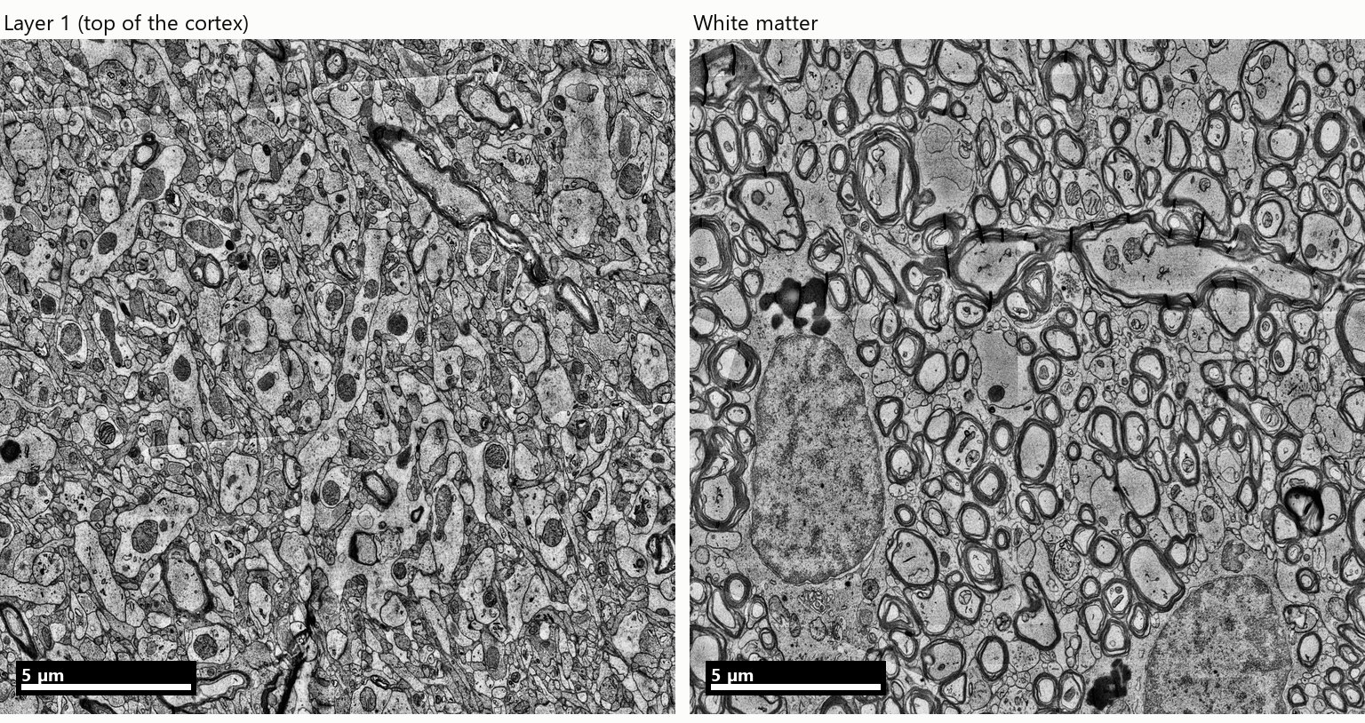
Raw H01 electron-microscope tiles from the same sample, 20 × 20 µm each. Left: layer 1. Right: white matter. Greyscale as imaged, not recoloured.
What you are looking at
Each tile is a single 20 × 20 µm view from the raw H01 images, one placed at the middle of the layer-1 cells and one at the middle of the white-matter cells (the median cell position in each, from the release's somas.csv table).
- Layer 1 is a dense tangle of thin processes, dendrites, unwrapped axons and the branches of support cells. Almost none of them carry a myelin ring.
- The white matter is mostly round profiles, each ringed by a thick dark band. These are axons cut across, and the dark band is the myelin sheath. The H01 authors note that "the myelin in the white matter ran primarily orthogonal to the plane of the section", which is why we see the fibres end-on as rings. The two large grainy ovals are cell nuclei.
The colours are reversed in these images. The tissue was stained with heavy metals (osmium and uranyl acetate) before imaging, and myelin comes out dark. "White" describes how the tissue looks to the naked eye.
The number, layer by layer
The H01 team assigned each reconstructed piece of tissue to a category (unmyelinated axon, myelinated axon, dendrite, astrocyte, cell body and a few smaller ones, plus blood vessels) and reported what share of each layer's volume each category fills (Table S4). The myelinated-axon share:
| Layer | 1 | 2 | 3 | 4 | 5 | 6 | White matter |
|---|---|---|---|---|---|---|---|
| Myelinated axons, % of volume | 1.0 | 0.8 | 2.5 | 4.8 | 5.6 | 7.7 | 31.5 |
In the white matter, the rest is unmyelinated axons (27.9%), dendrites (16.5%), astrocytes (15.4%), cell bodies (6.2%) and blood vessels (2.6%).
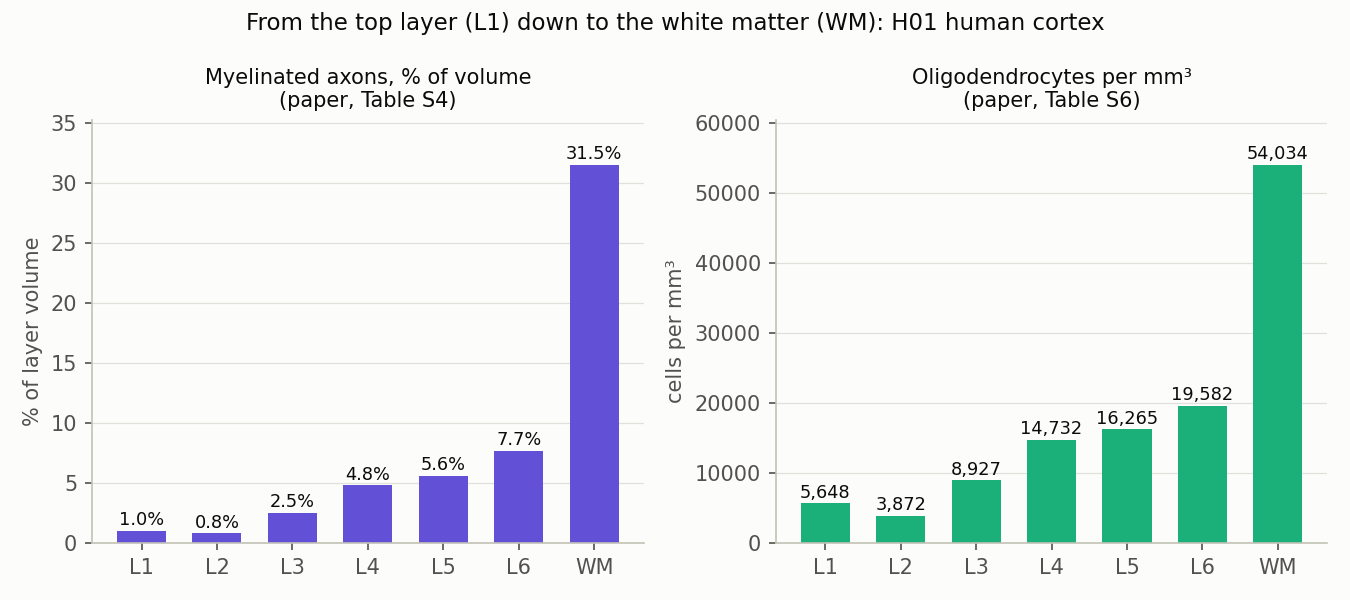
Left: myelinated-axon volume share. Values: H01 paper, Table S4. Right: oligodendrocyte density, corrected for tissue compression. Values: H01 paper, Table S6. These are the paper's numbers, not our measurements.
The two charts rise together, and that is expected. Myelin in the brain is made by oligodendrocytes, so the cells and their product follow the same gradient. The paper says so directly: "Myelin, a product of oligodendrocytes, followed the oligodendrocyte density gradient, with the highest density in the white matter and the lowest in layer 2." In our own count of the same sample, 77% of the cells in the white matter are oligodendrocytes (see What is the most common cell in the human brain?).
Read the 31.5% carefully
Two details in the paper's method change what the number means:
- The sheath itself is not counted. The category is "myelinated axons excluding their myelin sheaths": it measures the inside of the wrapped axons, not the myelin around them.
- Part of the tissue is left out. The analysis "excluded 36.13% of the voxels" in the imaged tissue, for example myelin sheaths, tears in the tissue, image defects and the space between cells.
So 31.5% is the share of the classified white-matter tissue that is the inside of myelinated axons. If the sheaths were added back, myelin-wrapped fibres would make up an even larger share of that tissue. It is not the share of every cubic micrometre, and it is not a measure of how white the tissue looks.
Why bother wrapping axons?
Speed. An oligodendrocyte wraps many layers of its own membrane around a stretch of axon. The wrapped stretches are separated by short bare gaps, the nodes of Ranvier, and the electrical signal effectively jumps from gap to gap. This is called saltatory conduction, and it is much faster than conduction along a bare axon of the same size (Stadelmann et al. 2019). The same review notes that myelin and oligodendrocytes do more than this, including supporting the axon's metabolism.
White matter is where the brain's long-distance cables run, connecting cortical areas to each other and to deeper structures. Fast cables for long distances is the trade it makes, and the fat-rich wrapping is why that tissue looks white.
The map we used
H01 is a fragment of human temporal cortex, removed during epilepsy surgery, imaged with an electron microscope at nanometre resolution and reconstructed in 3D (Shapson-Coe et al. 2024, Science). The slab runs from layer 1 down into the white matter below the cortex, and it is public under CC BY 4.0. The images above come from the release's raw image volume at 8 nm per pixel, downscaled for the web.
What this does not show
- One sample. One person, one area of temporal cortex, removed during epilepsy surgery. The white matter in it is only the part the slab reached just below the cortex.
- The paper's values, not ours. We did not re-measure the volume shares. They depend on the paper's automated classification and on the 36% of voxels it left out.
- Two images are not a survey. Each tile is one 20 µm view. They show what the numbers describe, but they are examples, not the measurement.
- Colour was not measured. We did not look at this sample's colour. The link between myelin's fat and the white look comes from the references below, not from H01.
References
- Shapson-Coe A, Januszewski M, Berger DR, et al. A petavoxel fragment of human cerebral cortex reconstructed at nanoscale resolution. Science. 2024;384:eadk4858. doi:10.1126/science.adk4858
- O'Brien JS, Sampson EL. Lipid composition of the normal human brain: gray matter, white matter, and myelin. J Lipid Res. 1965;6:537–544. doi:10.1016/S0022-2275(20)39619-X
- Santos EN, Fields RD. Regulation of myelination by microglia. Sci Adv. 2021;7:eabk1131. doi:10.1126/sciadv.abk1131
- Stadelmann C, Timmler S, Barrantes-Freer A, Simons M. Myelin in the central nervous system: structure, function, and pathology. Physiol Rev. 2019;99:1381–1431. doi:10.1152/physrev.00031.2018
Related
What Is the Most Common Cell in the Human Brain? In This Sample, Not the Neuron
Nov 2 · 6 min read
NeuroscienceHow Close Are Neurons to Blood Vessels? We Measured 10,441 of Them in a Real Human Brain Sample
Oct 30 · 6 min read
NeuroscienceWhich H01 Is It? Two Public Synapse Releases, Only One Matches the Paper — and How We Found Out the Hard Way
Oct 19 · 8 min read
NeuroscienceWe Followed One Neuron Through a Map of the Human Brain — Methods, Numbers, and What We Got Wrong
Oct 1 · 9 min read