What Is the Most Common Cell in the Human Brain? In This Sample, Not the Neuron
In about one cubic millimetre of human cortex mapped with an electron microscope (H01), oligodendrocytes outnumber neurons 20,139 to 16,087. Half of them sit in the white matter. In the grey matter alone, neurons still win. Here is the count, layer by layer.
This is the companion post to our Short The most common cell here isn't a neuron. The numbers are also on our Numbers page, in two cards: oligodendrocytes vs neurons and white-matter oligodendrocytes.
The short answer
In one real piece of human cortex, about one cubic millimetre, the most common cell is not the neuron. It is the oligodendrocyte, the cell that wraps nerve fibres in myelin. We counted 20,139 oligodendrocytes and 16,087 neurons (all neuron types added together). The H01 authors say the same thing in their abstract: "oligodendrocytes were the most common cell".
There is a catch, and it matters. The sample reaches down into the white matter under the cortex, and half of all its oligodendrocytes (10,110) sit there. If you count only the grey matter (layers 1 to 6), neurons outnumber oligodendrocytes, about 15,000 to 9,400. So the honest answer is: in this sample, yes; in the grey matter alone, no.
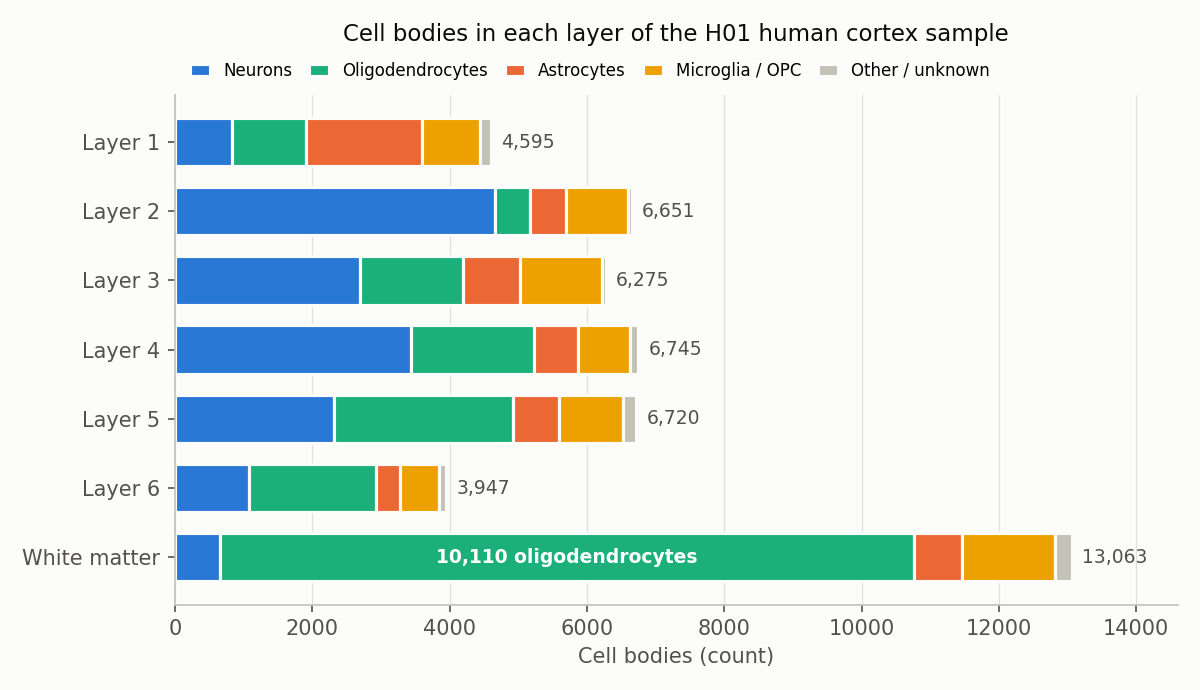
Cell bodies in each layer, by type. Our count from H01 somas.csv, using the dataset's own cell-type and layer labels. 1,383 cells have no layer label and are not shown.
What about the whole brain?
One sample cannot answer that, and it is worth saying what is known. The best-known whole-brain count, by Azevedo et al. (2009), found about 86.1 billion neurons and 84.6 billion non-neuronal cells in the adult male human brain: close to one to one, not the "ten glia for every neuron" that is often quoted. Their method separates neurons from everything else, so it does not tell you which single non-neuronal type is the most common across the whole brain. Our sample is a much smaller window, but it shows the same pattern from the inside: the answer depends on how much white matter you include.
The map we used
H01 is a fragment of human temporal cortex, removed during epilepsy surgery, imaged with an electron microscope at nanometre resolution and reconstructed in 3D (Shapson-Coe et al. 2024, Science). It spans all six cortical layers plus some of the white matter below them, and it is public under CC BY 4.0.
The release includes a table, somas.csv, with one row per cell body: its position, its cell type and its layer. The H01 team labelled the cell types by hand, "based on their appearance in the EM sections and their 3D shape", and assigned layers by clustering the cell bodies by position and size.
How we counted
- Cell types. We grouped five labels as neurons: pyramidal, interneuron, spiny atypical, spiny stellate and unclassified neuron. That gives 16,087. Oligodendrocytes are a single label: 20,139. Both numbers match the paper's Table S5 exactly, as do astrocytes (5,474) and microglia with their precursor cells, OPCs (6,536).
- Layers. We split the counts by the table's own layer label. 1,383 cells have no layer label (434 neurons and 679 oligodendrocytes among them), so the grey-matter and white-matter counts do not add up to the totals.
- Grey vs white. Layers 1 to 6 hold 15,005 neurons and 9,350 oligodendrocytes. The white matter holds 13,063 cells, and 77% of them (10,110) are oligodendrocytes. Only 648 are neurons.
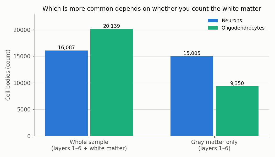
Neurons vs oligodendrocytes, with and without the white matter. Our count from H01 somas.csv.
Layer by layer
The mix of cells changes a lot with depth:
- Layer 1, the thin top layer, has few neurons. Its most common cell is the astrocyte (1,688 of 4,595 cells).
- Layers 2 to 4 are neuron territory. Pyramidal neurons are the most common single type in each.
- Layers 5 and 6 tip the other way. Oligodendrocytes are the most common type, at 39% and 47% of cells.
- The white matter is 77% oligodendrocytes.
So oligodendrocytes become more common the deeper you go. The H01 authors describe the same gradient and the reason for it: oligodendrocytes "were distributed according to a gradient, with the lowest density in the upper layers and highest density in the white matter, as expected, given their role in myelin formation."
One more detail if you split neurons into their types: even in the grey matter, oligodendrocytes (9,350) outnumber pyramidal neurons (8,302), the largest single neuron type. Whether the oligodendrocyte "wins" depends on whether you lump all neurons together.
Why the white matter is full of them
Oligodendrocytes make myelin, the fatty, many-layered wrapping around axons in the brain and spinal cord. Myelin lets electrical signals travel along an axon much faster (Stadelmann et al. 2019). White matter is mostly long, myelin-wrapped axons running between brain areas, so that is where the myelin makers gather. We look at that wrapping directly, in electron-microscope images from the same sample, in Why is white matter white?
Glia vs neurons in the same sample
Oligodendrocytes are one kind of glia, the brain's non-neuronal support cells. Counting all glia (astrocytes, oligodendrocytes, microglia and OPCs), the paper reports that "glia outnumber neurons 2:1 (32,315 versus 16,087)". That ratio also includes the white matter. In layers 1 to 6 alone, our count gives about 1.3 glia per neuron (19,238 vs 15,005).
What this does not show
- One sample. One person, one area of temporal cortex, removed during epilepsy surgery. The authors note they cannot rule out that long-term epilepsy or its treatment had subtle effects on the tissue.
- How much white matter was cut. The whole-sample ratio depends on how deep the surgical piece and the imaged slab happened to reach. A sample with more white matter would give an even bigger oligodendrocyte lead; one with none would give a neuron lead.
- The dataset's own labels. Cell types were assigned by people looking at the images, and layers by a clustering method. We did not re-check either.
- Counts, not size. This is a count of cell bodies. Neurons are larger and their branches fill much more of the tissue, so "most common" does not mean "takes up the most space".
Code and data
- Cell bodies: H01
somas.csv(positions, cell types and layers; 49,379 rows). - Counting: a short Python script that tallies the
celltypecolumn by thelayercolumn. The two figures are generated by the same script. - Cross-checks: paper Table S5 (total counts) and main text.
References
- Shapson-Coe A, Januszewski M, Berger DR, et al. A petavoxel fragment of human cerebral cortex reconstructed at nanoscale resolution. Science. 2024;384:eadk4858. doi:10.1126/science.adk4858
- Azevedo FA, Carvalho LR, Grinberg LT, et al. Equal numbers of neuronal and nonneuronal cells make the human brain an isometrically scaled-up primate brain. J Comp Neurol. 2009;513:532–541. doi:10.1002/cne.21974
- Stadelmann C, Timmler S, Barrantes-Freer A, Simons M. Myelin in the central nervous system: structure, function, and pathology. Physiol Rev. 2019;99:1381–1431. doi:10.1152/physrev.00031.2018
Related
Why Is White Matter White? 31.5% of Its Mapped Volume Is Myelinated Axons
Nov 2 · 7 min read
NeuroscienceHow Close Are Neurons to Blood Vessels? We Measured 10,441 of Them in a Real Human Brain Sample
Oct 30 · 6 min read
NeuroscienceWhich H01 Is It? Two Public Synapse Releases, Only One Matches the Paper — and How We Found Out the Hard Way
Oct 19 · 8 min read
NeuroscienceWe Followed One Neuron Through a Map of the Human Brain — Methods, Numbers, and What We Got Wrong
Oct 1 · 9 min read